
These cases are presented as a product of my partnership with Dr Vicki Bowes, Avian Vet/Pathologist who very kindly offered to help me decipher the chicken health mysteries in my ‘What Is This?’ file. Some contain necropsy photos of deceased birds, while others have happier endings. In either case, I hope they provide you with useful information on potential illnesses and what you might look out for or do to avoid them.
“I had a chicken that I was treating for a crop impaction. Unfortunately she died before I could take her to the vet.
She was skin and bones from not getting proper nutrition and had a pretty large impaction. I decided to perform a necropsy to see what was causing the impaction and found some other very interesting things.
She had these white/yellow spots in her abdomen and they appeared to have been growing mold on them. She had only been dead for 4 hours so she had to have been living with this condition. I also found: one of her lungs to be possibly necrotic or having some of these deposits on them; blackened mesentery/bowel; a mass on her liver and her crop appeared to be ulcerated.” – Brittany Myers
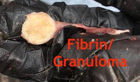
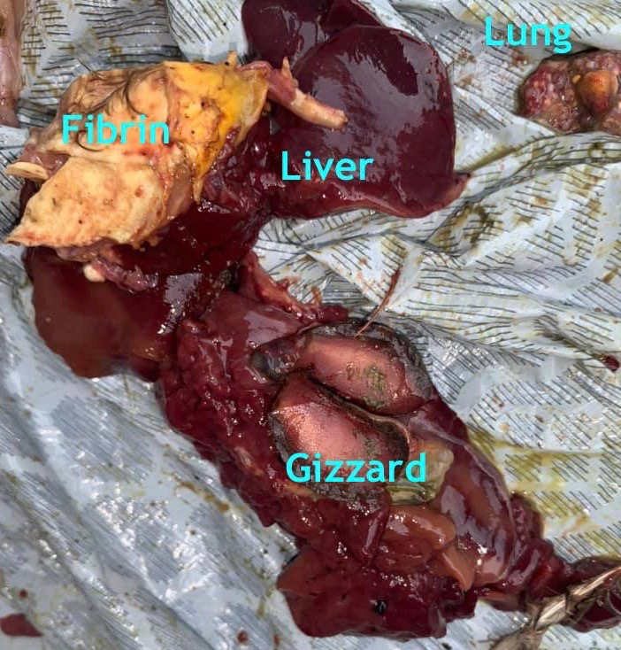
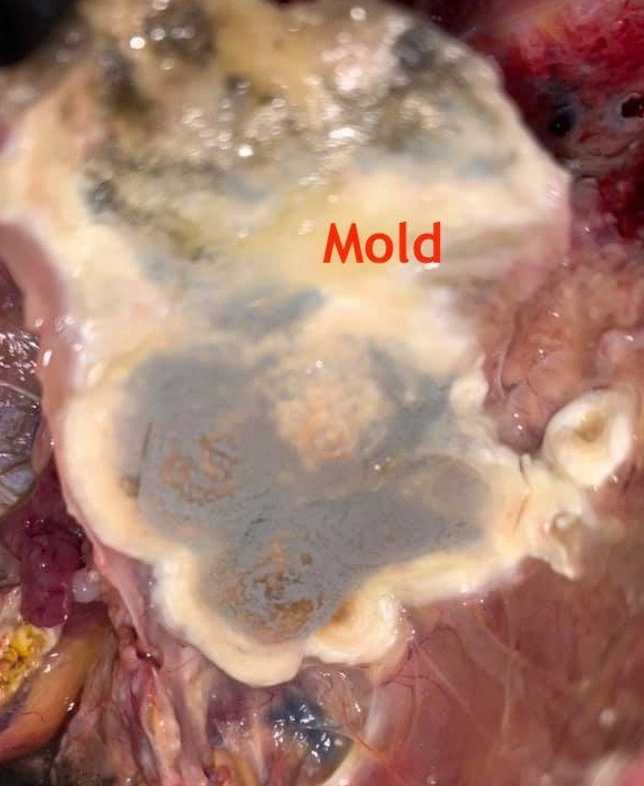
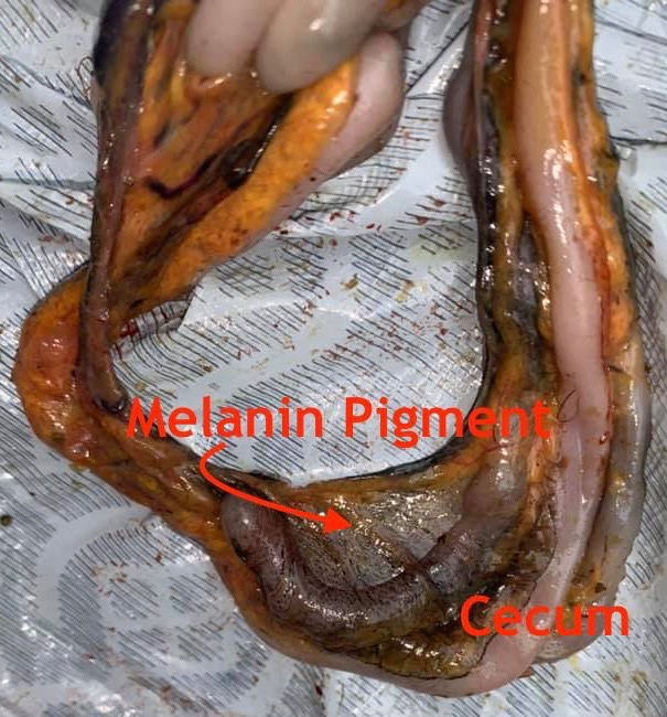
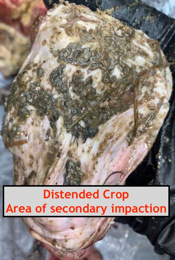
Looking at these DIY necropsy photos one might be reminded of a moldy piece of bread. Lots of us have heard of the health risks of black mold in our homes. Unfortunately all those dark patches are indicative of a fungal infection inside the bird’s body.
Dr Bowes: The distended crop reported by the owner is a secondary impaction. The fungal infection was caused by spores ingested or inhaled by the bird coming into contact with spilled wet feed or bedding. The progression of their spread occurs slowly, affecting the bird’s respiratory and other systems. The yellow fibrin is an indicative of the inflammatory process.
Prevention measures would be to reduce opportunities for birds to eat wet feed and to clean your coop regularly, especially in wet or cold weather. Never give your birds moldy food.
Necrotic Foot
“My chicken’s foot just fell off… or she ate it off. What can I do? She was already in the house with bumblefoot. I think my hubby squeezed her foot too hard trying to get the infection out. She stopped walking on it and just held it up, and then I found her foot when cleaning her cage this morning. But her other foot doesn’t look right either.” – Jenn Stone
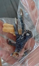


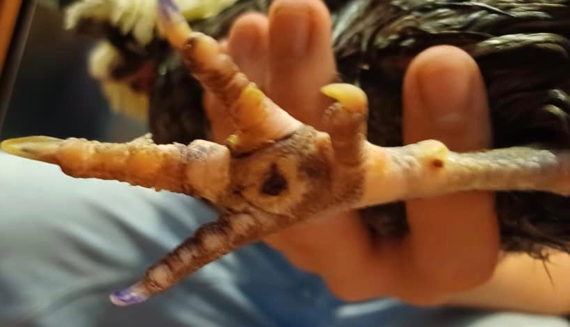
Dr Bowes: What may have started as a small bumblefoot lesion caused by a bacterial infection, then became a chronic issue that spread from the foot up the leg. An untreated infection can lead to swelling, damage to blood vessels and circulation that is compromised so the affected area loses blood flow. In these photos, the stump is already healed and rounded and the blackened area is necrotic, meaning the foot was dead long before it fell off.
Unfortunately in this case the good foot also has bumblefoot. Dr Bowes suggested that if the bird had one leg that was capable of bearing weight it might survive, but given its current condition it would struggle, the foot would become worse and the stump would become ulcerated. Her recommendation was humane euthanasia.
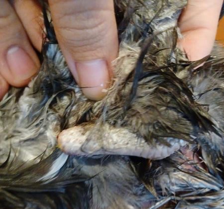
“Madonna had a large infection in her sinus just in front of her eye. We laid her on the side so she was flat on the ground. We put anesthetic ointment on the area and were able to cut the whole mass out. She didn’t react at all during the surgery. We used strong surgical tape to close the incision and she made a full recovery.” – Joel Edahl
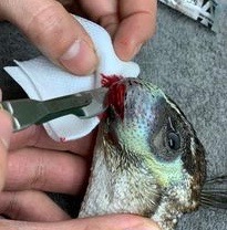
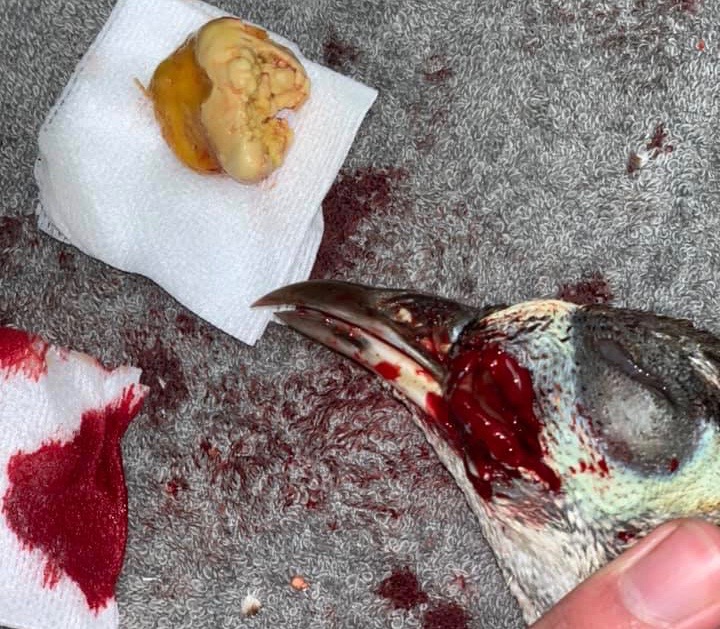
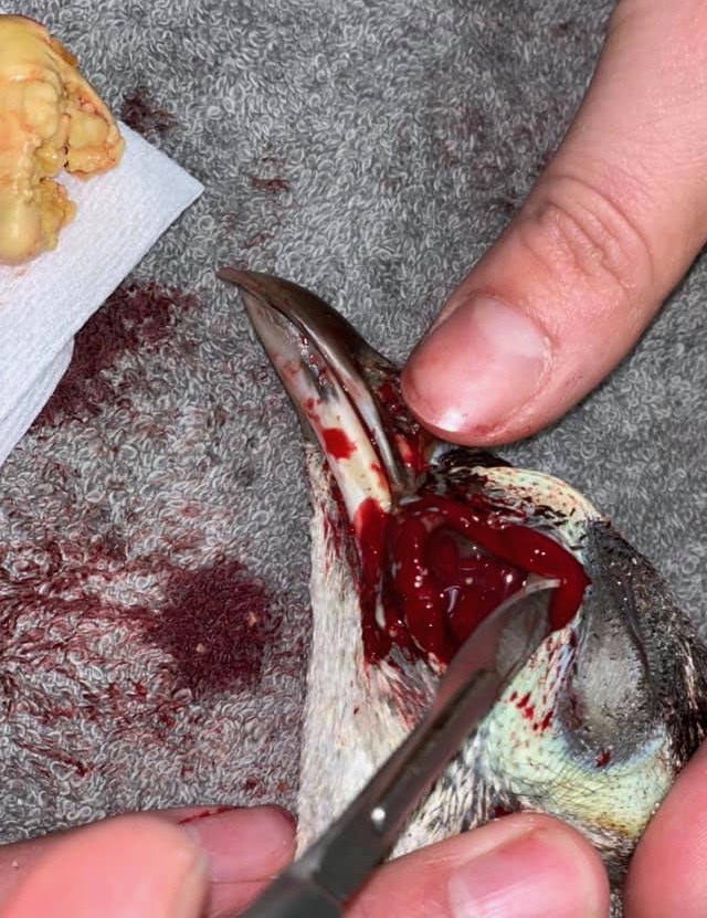
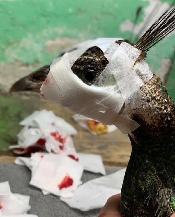
This is the first peafowl I’ve featured, but the infection is common in poultry as well. Dr Bowes believed it was a bacterial infection, either Staphylococcus, Pseudomonis or Mycoplasma gallisepticum. If the latter, the bird would be a carrier for life.
Of course, she probably wouldn’t have recommended at-home surgery, but veterinary care is not always available or affordable and a bird would be euthanized. In this case, the owners did a good job of excising the caseous (cheesy) material from the intraorbital sinus, then cleaning the area and wrapping it. Dr Bowes suggested rinsing the wound with an antibacterial flush, giving oral or injectable antibiotics and monitoring the site for infection.
Meat Bird Necropsy
“I processed a failure to thrive Cornish x 8 week old broiler today. She had half a dozen hard pale growths inside the body cavity and some yellow gelatinous nodes near her liver. Does this look like cancer or something else? We processed a dozen from the same batch yesterday and didn’t see any like this in those. Meat was not consumed.” – Sarah Violet Atkinson
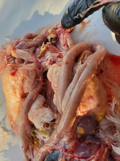
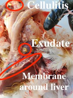
Various breeds of meat birds have been developed to put on lots of weight in a short period of time. Unfortunately their lives until butcher date is quite short, often 6-8 weeks! They are prone to a number of health issues related to their rapid weight gain as well as vulnerabilities to bacterial infections.
As soon as Dr Bowes saw these DIY necropsy photos she diagnosed it as an E.coli infection, common in broilers. Chicks can become infected through the yolk sac or navel at hatch. Infectious Bronchitis Virus can also introduce E.coli into the body through a respiratory infection. Some birds die quickly, while others may do well for three or four weeks before become symptomatic.
If you have a chicken health issue you’d like solved drop me a line using the contact button on the home page.

Gross, but very informative. Thanks.
LikeLiked by 1 person
Poor Madonna! Kudos to her owners for being willing to try anything. I’m assuming vet care was not possible. Bless her little heart
LikeLiked by 1 person