
For the last three decades I’ve worked doing education of some kind in the field of HIV, Hepatitis C and harm reduction. I’ve mentored practicum students and peers. No matter how much I know, I always have more to learn and nothing pleases me more than a relationship of reciprocal learning.
Several months ago Dr Vicki Bowes, Avian Vet/Pathologist from the Animal Health Centre offered to meet up with me on a regular basis to help me figure out the unsolved mysteries in my ‘What Is This?” file. We’ve been going through case notes, necropsy reports and photos to reach what Dr Bowes terms her ‘best guess’.
We recently spent a marathon 4½ hours sifting through my files and I was so thrilled when I presented her with some health issues she’d never even heard of. It was a great opportunity to teach my mentor something new and feel like our relationship is a bit of a two-way street. So read on if you’re interested in seeing what tricks two old dogs can both learn and teach.
Cutaneous Horn (Cornu cutaneum)
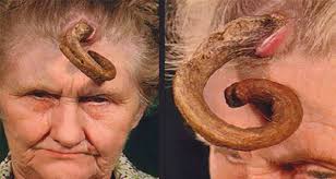

When I first showed Dr Bowes this chicken photo (credit: Jeni Barker) she had no idea what she was looking at. The scientific term is conical hyperkeratosis cutaneous horn, which is a mouthful meaning a lesion consisting of keratotic material above the skin that resembles an animal horn.
Keratin, of course, is a type of protein that makes hair, fingernails, claws and feathers. Cutaneous horns can be found in a number of species including cats, dogs, people and chickens. The horns come in various shapes and sizes and are often benign; in people, they can be linked to skin cancer. They can be removed, but often regrow.
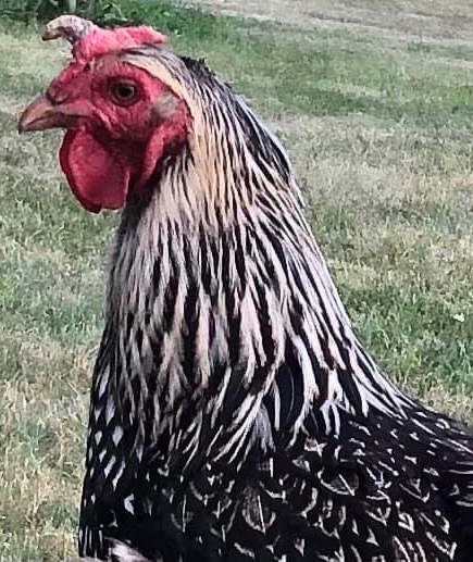
Herniated Tongue

I’ve seen several posts like this in which folks suggest that the protrusion is the bird’s beak that has come through the skin under the chin. Dr Bowes wasn’t convinced of this theory and was itching for more definitive photos and notes. Neither case was accompanied by photos of the inside of the mouth, so we couldn’t see if the tongue was actually in the correct position or not. Was the bird able to function? Was the tongue moving? Was there a clear exit point under the chin? How long did these birds survive like that? Inquiring minds want to know, Dr Bowes most of all. So much so, she’s prepared to offer a free necropsy on any bird sent to her suffering with this condition.
Ectopic Growths
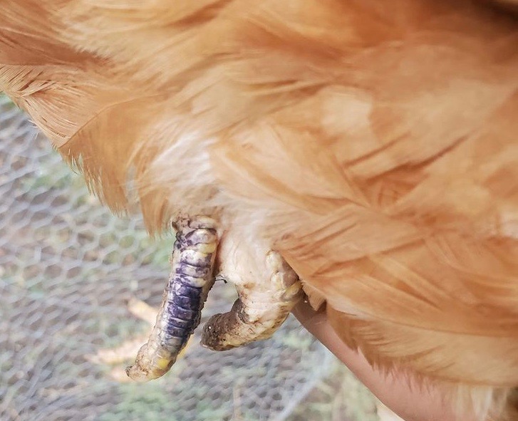
A vestigial twin occurs when one organism is normal and the other, parasitic twin is malformed or incomplete, usually consisting entirely of extra limbs or organs. Most vestigial limbs are non-functional, and although they may have bones, muscles and nerve endings, they aren’t part of, or under the control of, the host.
I presented this photo as vestigial toes, but Dr Bowes suggested it was more likely to be ectopic rather than embryonic tissue. That means the cells came from the host, but grew in the wrong place. I had a hen that as an adult grew what appeared to be one long, skinny toe on each of her legs, complete with scales, but totally non-functioning as toes.
Personally I’m into the oddities and necropsies. I find posting those kinds of cases and photos is really polarizing: you either love them or hate them. This one will probably be no different. If you’re in the latter camp, look away fast.
Mucoceles
I snagged the following post from an online group. This comment was made from a veterinarian advisor in response to seeing the photo on the left: “The esophagus and crop are made of similar tissue compared to the colon/vent. Pressure and straining can cause prolapse. I don’t know what caused this crop to prolapse – trauma, squeezing, being held upside down, poor anatomy, crop impaction, etc. There are too many factors involved to narrow it down to just one.” (Name Withheld)
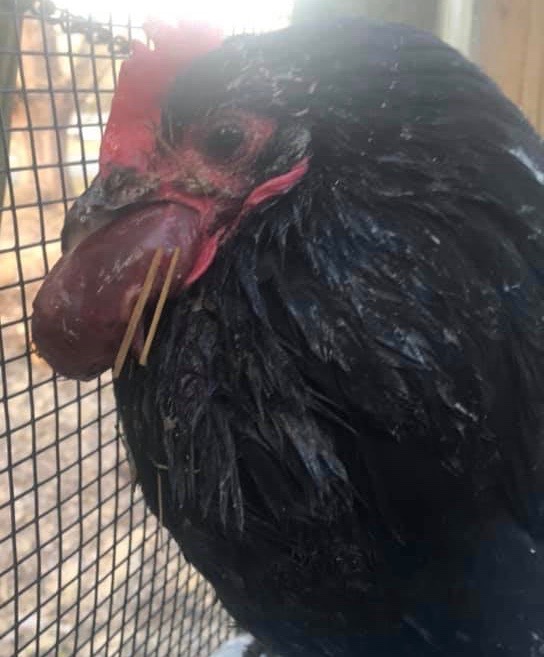
When I met up with Dr Bowes last summer I asked her about prolapsed crop and she’d never heard of it.
I sent her that photo and the vet’s comments and this was her response: “My first, and final reaction to the “prolapsed crop” is ABSOLUTELY NOT. The mass does not have the tissue consistency or conformation of a crop. For a crop to prolapse (anatomically next to impossible, it’s tacked down pretty tightly to adjoining tissue unlike an oviduct or rectum), it needs to evert so that the lining would be exposed to the outside and that certainly is not the case here.”
“A key piece of the puzzle would be the duration and final outcome. My best educated guess is that this is a tumour, possibly of muscle. I’ve seen tumours involving the supportive structures of the larynx. At this point I would have loved to touch it and biopsy it to get a definitive answer, but I’m pretty sure it’s not crop.”
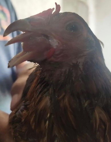
Over the next five months I saw two more cases posted in online groups, one of them attributed to being caused by a snakebite and the other, a wasp sting. Since this is clearly a ‘thing’ with chickens we recently took a closer look at the photos, some of which were accompanied with notes.
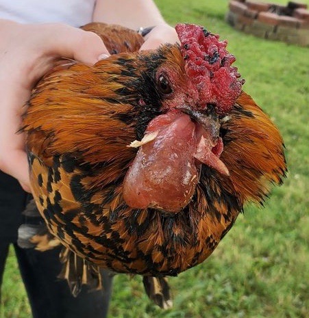
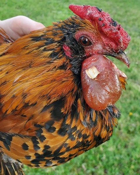
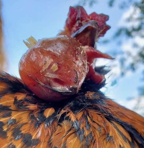
In all three cases, the birds presented as normal at the beginning of the day and then, within hours, had a fleshy mass protruding from their mouth.
After a little online reading Dr Bowes’s best guess was that this phenomenon is caused by trauma to the salivary gland, resulting in what is called a mucocele. For whatever reason, the gland gets impacted and a profuse amount of saliva builds up in short period of time expanding it under the tongue. She thought that if you tried to drain the fluid with a needle and syringe it would be a short-term fix because birds create lots of saliva as a way of digesting food in the absence of teeth to chew it.
I’m familiar with mucoceles as my dog had one – much smaller – under his tongue. My vet figured that a splinter from a stick might have damaged one of his saliva glands. He lived with it for well over a year, but when it started to get a bit bigger and redder from being abraded, the vet performed a procedure to deal with it.
In the case of mucoceles in chickens Dr Bowes made the following suggestions: a) take good photos, including inside the mouth, if possible, b) euthanize the bird immediately and c) send it to her lab and she would perform the necropsy free of charge just to be able see a case in person.
Update: February 2023: I came across a case that was successfully treated with the hen making a full recovery within days. Read about that case here.
Once again, my thanks go out to Dr Vicki Bowes, pathologist extraordinaire, for her expertise and generously offering to share that knowledge in our mutual pursuit of creating higher standards for small flock health outcomes.
If you have a mystery you’d like solved drop me a line using the contact button on the home page.
Featured photo credit: Dr Vicki Bowes

Woah….you certainly missed your calling!
LikeLiked by 1 person
oh I think a Hyperkeratosis cutaneous horn is what my chicken has on her foot. I was worried it was bumble foot.
LikeLiked by 1 person
Can you send me a photo? If you use the ‘contact’ button on my homepage I’ll send you my email.
LikeLike
plz take a look at these pics. Link found below.
Thx
https://m.facebook.com/groups/BackyardChickensBYC/permalink/10163045423709734/?ref=share&mibextid=NOb6eG
LikeLike
I’m not able to access the link you sent. Can you drop me a line using the ‘contact’ button on my homepage and I’ll send you an email where you can send the link or photos? Thanks
LikeLike