For more than three years I’ve gotten together with Dr Vicki Bowes, vet/ avian pathologist on a regular basis to pore over files on my memory stick loaded with interesting chicken health issues that I’ve collected for her expert opinion. She refers to it as ‘Show and Tell’, ‘Best Guess’ or, more recently, ‘Gorefest’ and has done a good job at making diagnoses given the information we have at hand. Sometimes all we are provided with is a short paragraph from the chicken’s owner, other times nothing more than a photograph.
My job is to write them up to share with my readers as a form of skills building for small flock keepers.
We met up recently to look at almost 60 cases. I’ve attempted to curate them according to the area of the body affected. These ones are grouped together as they all involve internal issues.
Broiler Liver
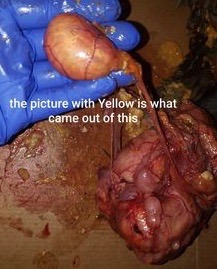
I was harvesting my broilers today and one came across a liver that I have never seen before. It’s firm and grey; the inside is red and almost pulverized. There is no odour, but actually smells rather pleasant. – Karlis Stegis

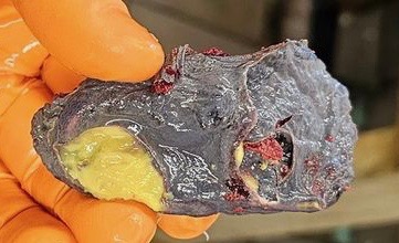
Dr Bowes: The yellow spot is fibrin. The liver is crumbly, friable, with a thickened capsule. It could be the result of septicemia (E.coli); mycoplasma from a respiratory infection like bronchitis; bacterial Hepatitis; or a hemorrhage. You would need to culture the sample to obtain an accurate diagnosis. Livers are a filter organ. If you are butchering birds that have diseased livers (or kidneys and spleens) dispose of the carcass and don’t eat it.
Worms & Marek’s Disease
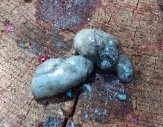
I unexpectedly lost a one-year-old Ayam Cemani rooster this morning. I was treating him for a hurt foot, expecting it to be bumblefoot. He ate fine last night while soaking in Epsom salt. I did give him two drops of Selenium and vitamin E in water with multivitamins. This morning, he had no energy and couldn’t stand up for long; he didn’t eat, then died. I didn’t get any feces this morning, so decided to do necropsy. I extracted what I could from his bowel and found a couple of worms and eggs. Is this coccidia or gapeworm eggs? The two example adult worms may not be identifiable. Not sure if one is torn or if that is a characteristic of a mouth/tail. I also found white spots on his liver.
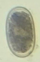


Dr Bowes: I would need to know the scale of these images to determine what kind of parasites we’re looking at. The first two are probably roundworm eggs; the third image is the internal structure of something that has been broken; the fourth might be a cecal worm. It’s unusual to see a whole worm in that field of magnification. I think the spots on the liver are tumours indicative of Marek’s Disease. If the sample had come from a turkey I might suggest ascarids which can wall themselves off and appear like those white spots.
Abnormal Testicle
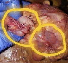
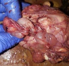
We processed a rooster today. I found this odd yellow patch on one of his testes. Can you tell me what this is? – Natalia Durazo


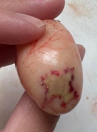
Dr Bowes: It’s an infarct caused by compromised blood circulation, like a blood clot. He could have lived normally with this issue.
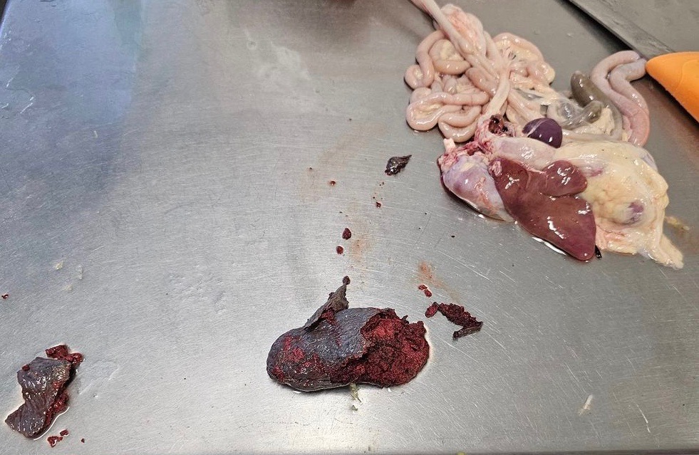
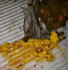
Bile (Michelle Edrees)
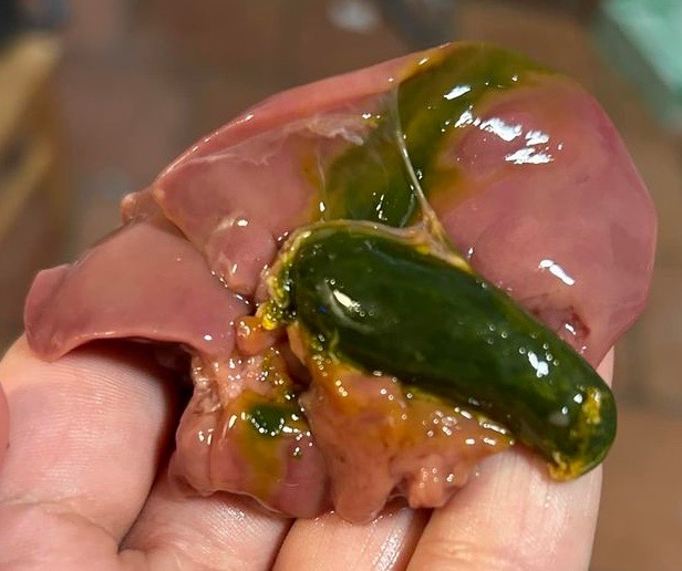
Dr Bowes: That’s a distended gall bladder, indicative of a bird that isn’t eating. The bile seeps into the adjacent tissue post-mortem. If you squeezed one end you would see it come out of the bile duct (not shown in photo). The liver looks normal. The cause of death can’t be determined by this photo and no accompanying information on symptoms.
Cystic Oviduct & Salpingitis (Darlenea Bingman)
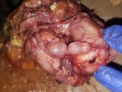
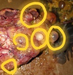
Dr Bowes: It’s difficult to tell from the photos what organs we’re looking at. It appears to be a cystic oviduct and salpingitis.
What is going on with my chicken? She presented with a head tilt yesterday and has not laid an egg in a week. She usually lays daily. I took her to work (dog and cat vet) for X-rays. I don’t know what the tiny circles are inside her either. – Kelsey McGuire
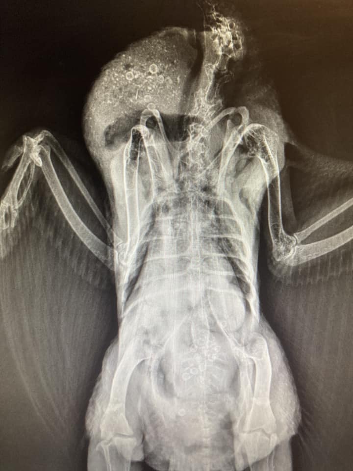
Dr Bowes: Judging from the shape and location I think those are cherry pits. They are not dark like lead that would result in neurological issues. I have no explanation for the head tilt.
Mystery Case
This is a 2-year old Bielefeld Kennhuhn hen. What is the sinuous thing on the X-ray? It was not visible on an X-ray taken nine months ago. The diagnosis I got was suspected Salpingitis. Prescribed: Suprelorin, calcium and Meloxicam (pain med). – Alex Nemetz
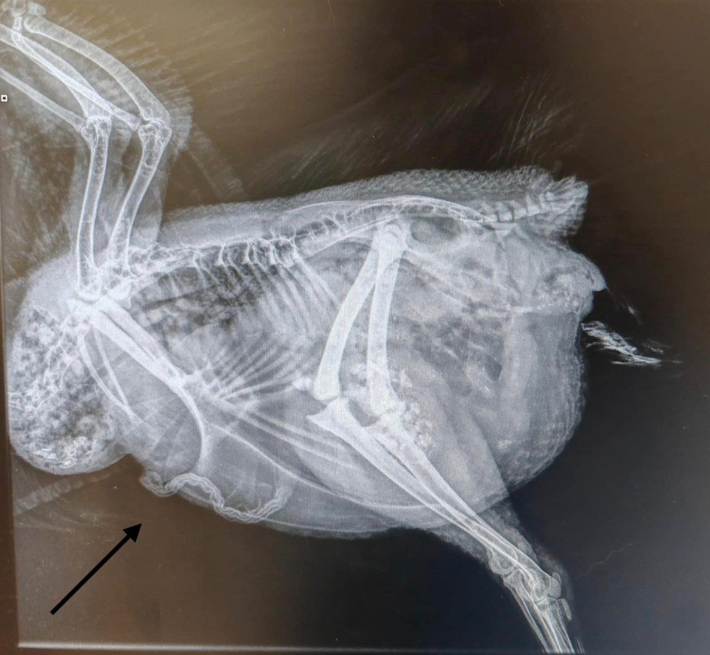
Dr Bowes: The object is dark, dense and has a structure. It is located outside of the sternum and under the skin. No worm would appear like that on a radiograph. I’m stumped.
Salpingitis
My vet said she’s not egg bound and couldn’t see fluid build up. She seems bloated, comb is a little purple, she’s still eating. – Kimberly Cortes


Dr Bowes: She is not egg bound. There is a tubular structure that I think is salpingitis (bacterial infection of the oviduct).
Silkie Kidneys
These are my 1½-year-old rooster’s kidneys. Why are they this gray and blueish? The rest of the organs were red. Just the kidneys were dark, so is it just a Silkie thing? – Autumndae Sampson
Bitchin’ Chickens: Purebred Silkies are fibromelanistic: they have black skin and dark internal organs.
Dr Bowes: Inflammation of the kidneys can result in swollen, congested dark red kidneys. Urates can change their colour to tan or white. I have never seen this blue-grey colour. Can you cut the kidneys so that a cross-section would reveal the internal colour?
Glossary
Fibrin: an insoluble protein formed from fibrinogen during the clotting of blood. It forms a fibrous mesh that impedes the flow of blood.
Fibromelanosis: a genetic mutation in some breeds of domestic chickens, expressed with abnormal accumulation of the dark pigment melanin in the skin and connective tissue
Infarct: a small localized area of dead tissue resulting from failure of blood supply
Suprelorin: a hormonal implant to stop a hen from laying to prevent/treat reproductive tract issues
Well, that wraps up another edition of Show & Tell With Bitchin’ Chickens and Dr Bowes. I hope that it’s been a learning experience for you.
If you’d like help with a case drop me a line using the ‘contact’ button on my home page. Remember to wear gloves, take good close up photos from several angles and supply us with plenty of information (e.g. timelines, symptoms, medications, general flock health, etc) so we’re able to more accurately pinpoint what’s going on.
Thanks again to Dr Vicki Bowes for her willingness to share her wealth of knowledge and experience to build capacity and skills in small flock keepers.
Featured photo credit: istock

Please tell me the Biefelder will be examined post mortem when her time comes!
LikeLike
Agreed. I wish more folks sent their birds for necropsy or, barring that, at least attempted to do a DIY necropsy and take some good photos. The mystery may continue.
LikeLike