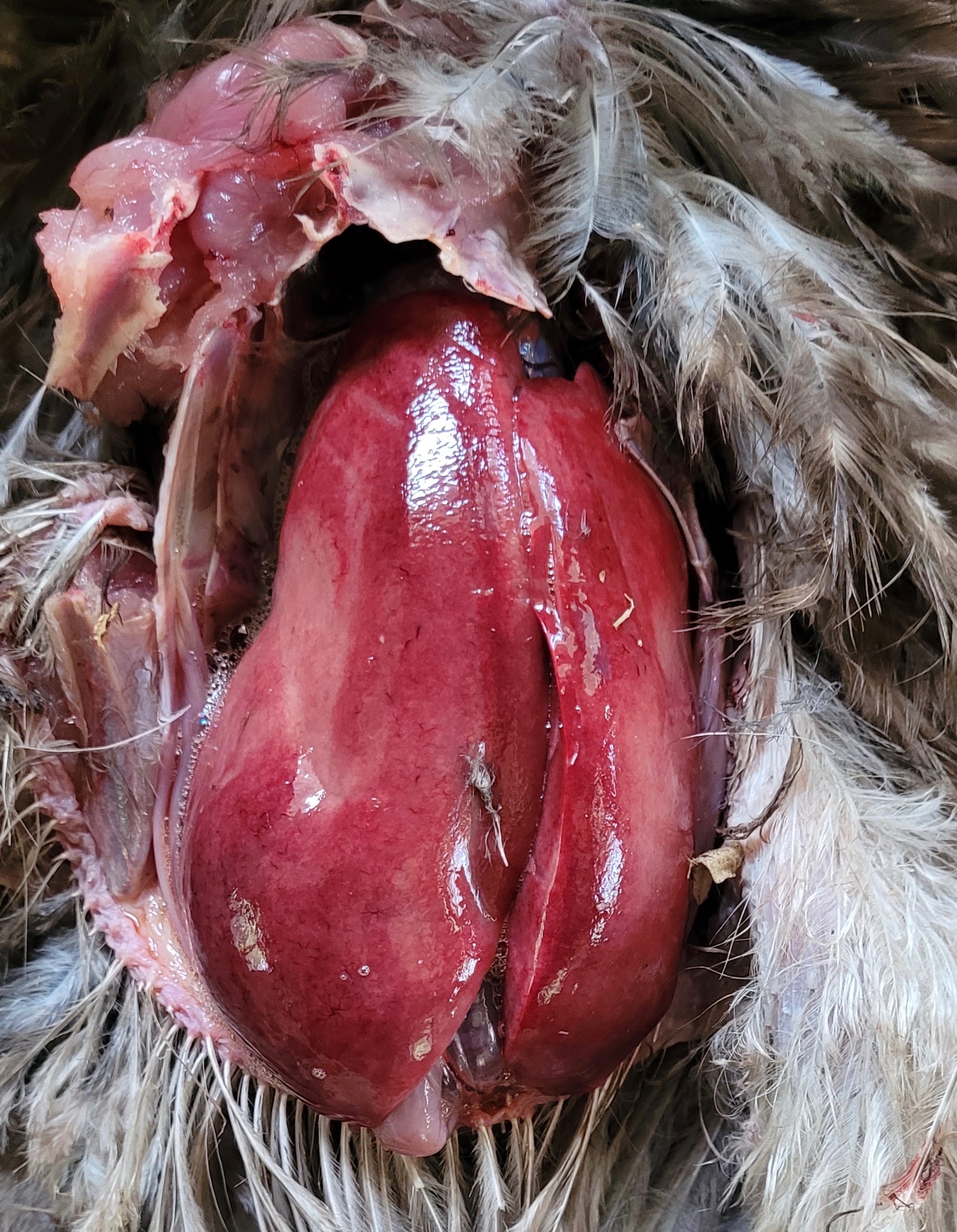
For more than three years I’ve gotten together with Dr Vicki Bowes, vet/ avian pathologist on a regular basis to pore over files in my memory stick loaded with interesting chicken health issues that I’ve collected for her expert opinion. She refers to it as ‘Show and Tell’, ‘Best Guess’ or, more recently, ‘Gorefest’ and has done a good job at making diagnoses given the information we have at hand. Sometimes all we are provided with is a short paragraph from the chicken’s owner, other times nothing more than a photograph.
My job is to write them up to share with my readers as a form of skills building for small flock keepers.
We met up recently to look at more almost 60 cases. I’ve attempted to curate them according to the area of the body affected. These ones are grouped together as they all involve DIY necropsies.
Brahma Hen, 2 years old (Jessica Beauchamp)
Symptoms:
- Found dead in the coop
- Heart had white layer over it
- Full crop
- Gall bladder appeared to have ruptured but I am not sure if that was post mortem
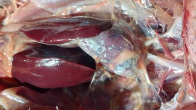
Dr Bowes: The white layer over the heart is urate deposits, a sign of dehydration. The green from the gall bladder is accumulated bile, indicative that she was not eating. The urates on the heart are typical of visceral gout. I suspect that she was deprived of water due to freezing conditions. It’s important that birds have access to water, regardless of the time of year.
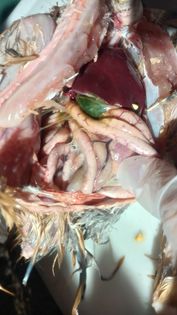
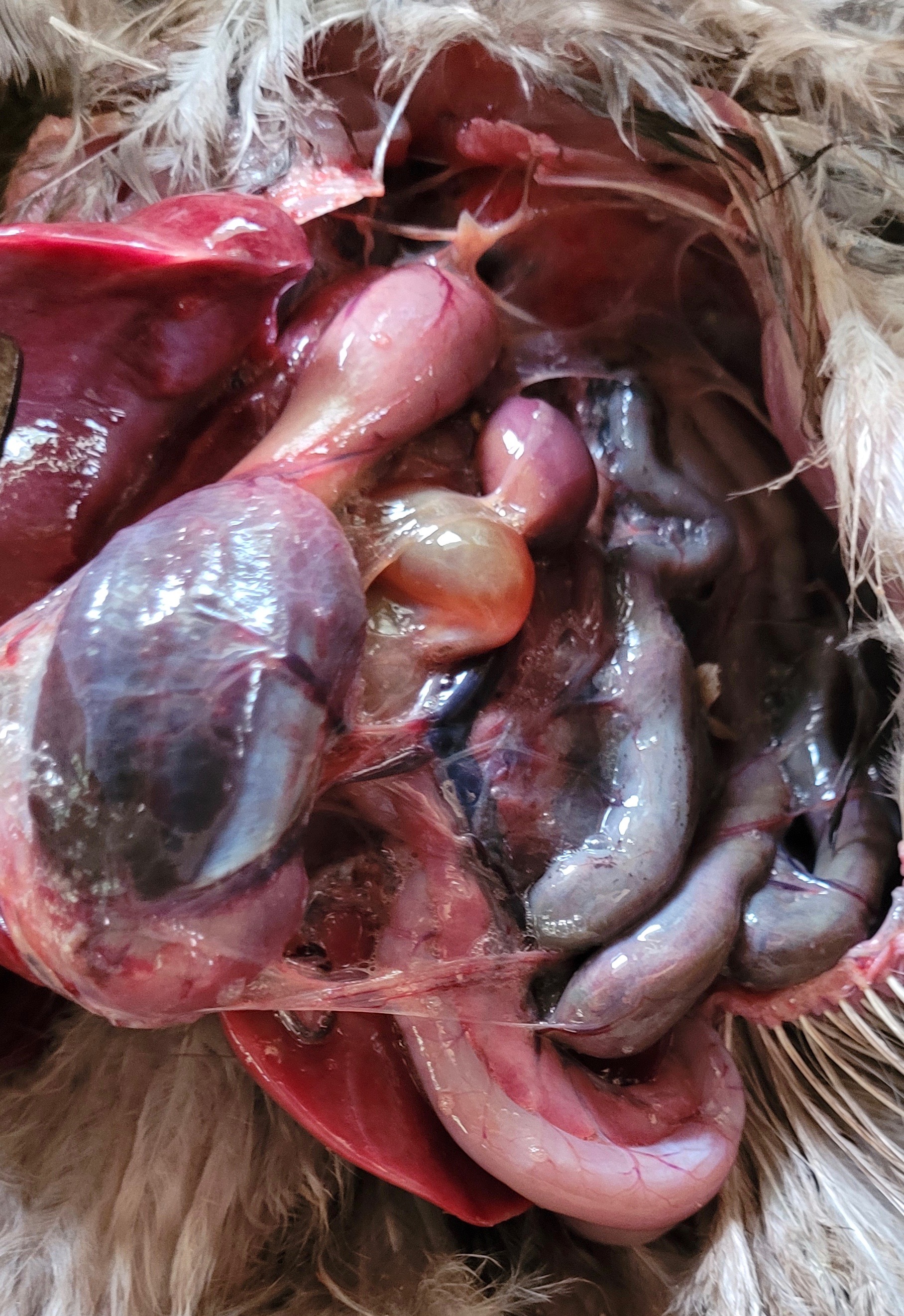
Leghorn Hen, 3 years old (Mindy Marin)
Symptoms:
- Showing signs of illness a few weeks ago
- Vent gleet and lethargy, then recovered
- Vent gleet returned
- Lethargy, not eating or drinking
Necropsy Findings:
- Food compacted in her mouth and sinuses
- Small fat deposits throughout her body
- Black tissue in thoracic cavity
- Dark, thin edges on liver
- Oviduct filled with tumor-like bumps and egg yolk
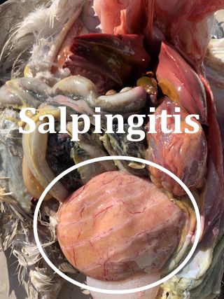
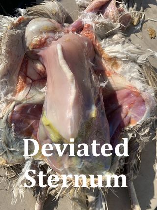
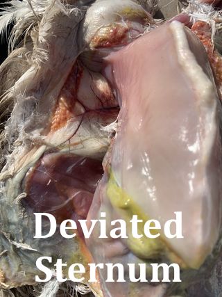
Dr Bowes: There is fibrin and peritonitis in her abdomen. The liver has crisp, normal margins. The black tissue is of no concern; it appears to be the result of a resolved hemorrhage or melanin. She has a sternal deviation, seen more commonly in high production layers as calcium gets leached out of bones to be used in egg production. The deformity can occur when there are high roost bars and hens experience impact upon crash landings on hard coop floors. The impaction in the groove of her mouth is just small feed. She has salpingitis (bacterial infection of the oviduct) and tumours. I would diagnose ovarian adenocarcinoma with secondary peritonitis.
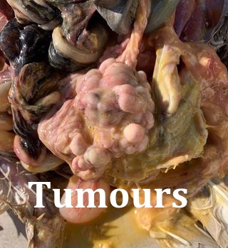
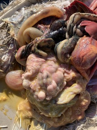
Barnyard mix, frizzle hen, 26 months old (Anonymous)
Symptoms:
- Lost back feathers over the summer due to rooster (who was rehomed two months ago), starting to grow back
- Comb was dark and not bright red
- Day prior to her death appeared slower, tucking head in, still eating
- Died overnight in the coop
Necropsy Findings:
- Prominent keel, poor body condition
- No evidence of egg production
- Empty crop
- Liver appeared large with tan areas, normal texture


Dr Bowes: She had a massively enlarged liver, marked with diffuse infiltration. There appears to be more tumour than actual liver. Serous atrophy with no fat stores, emaciation. The black pigment was normal melanosis. It is difficult to see if there were tumours in the spleen. Avian Leukosis Complex refers to either Leukosis or Marek’s Disease. In the lab tumour markers could be applied to microscopic tissues to look for T-cells which identify Marek’s Disease. Large immature cells are associated with Leukosis while mature cells are typical of Marek’s Disease. In this case, because of the hen’s age I would diagnose Leukosis (Marek’s usually affects young birds).
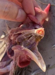
Appenzeller Spitzhauben x Rooster, 4.5 years old (Anonymous)
Symptoms: Day 1 – 3
- Open beak, drooling
- Matted wet feathers
- Head shaking, scratching face
- Squeaking sound
- Small lump in throat
- Empty crop
- Long strands of saliva
- Smell inside beak
- Small watery green poop
- Euthanized
Necropsy Findings
- Golf ball sized discrete lump in throat
- Yellow plaques at back of throat
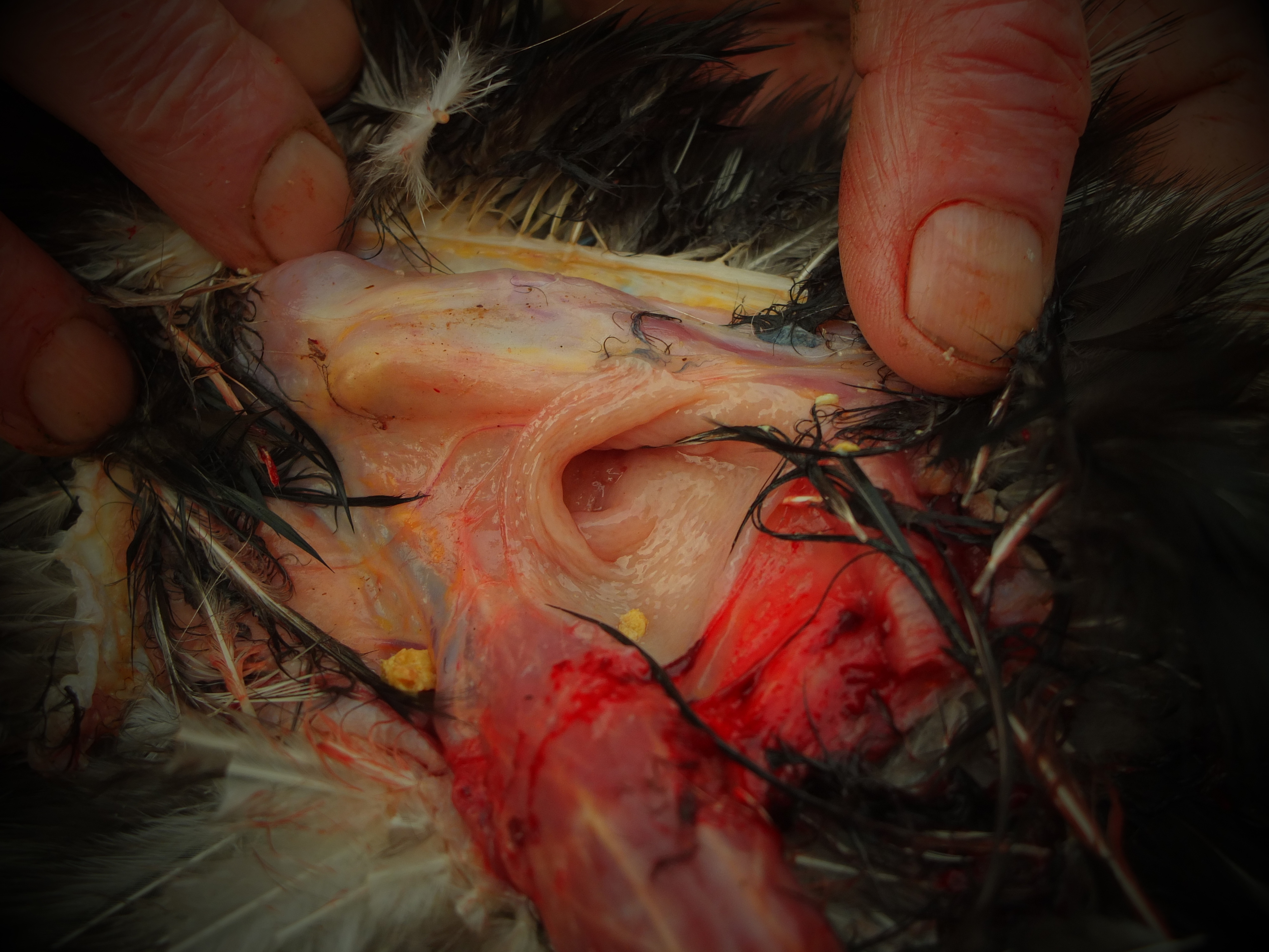
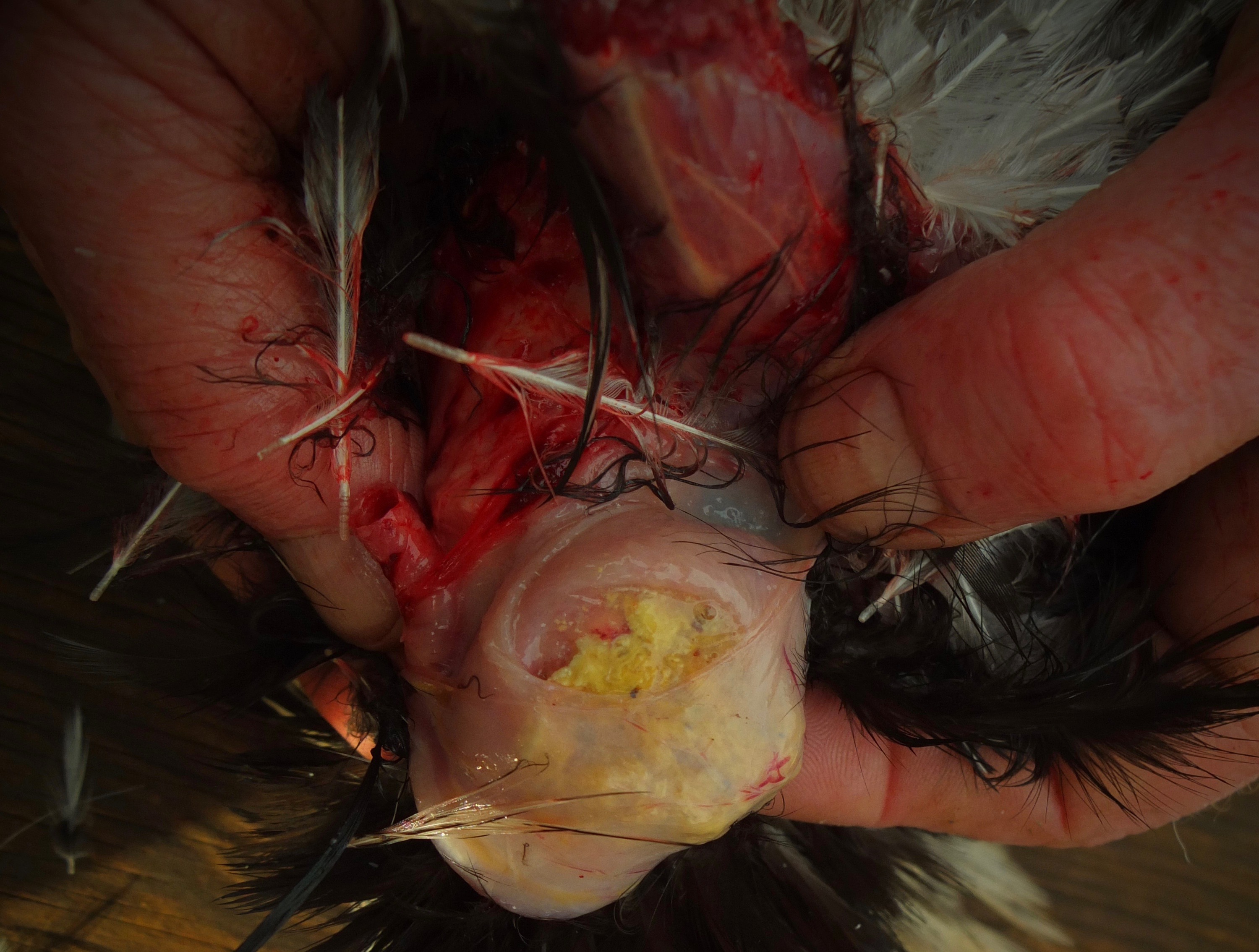
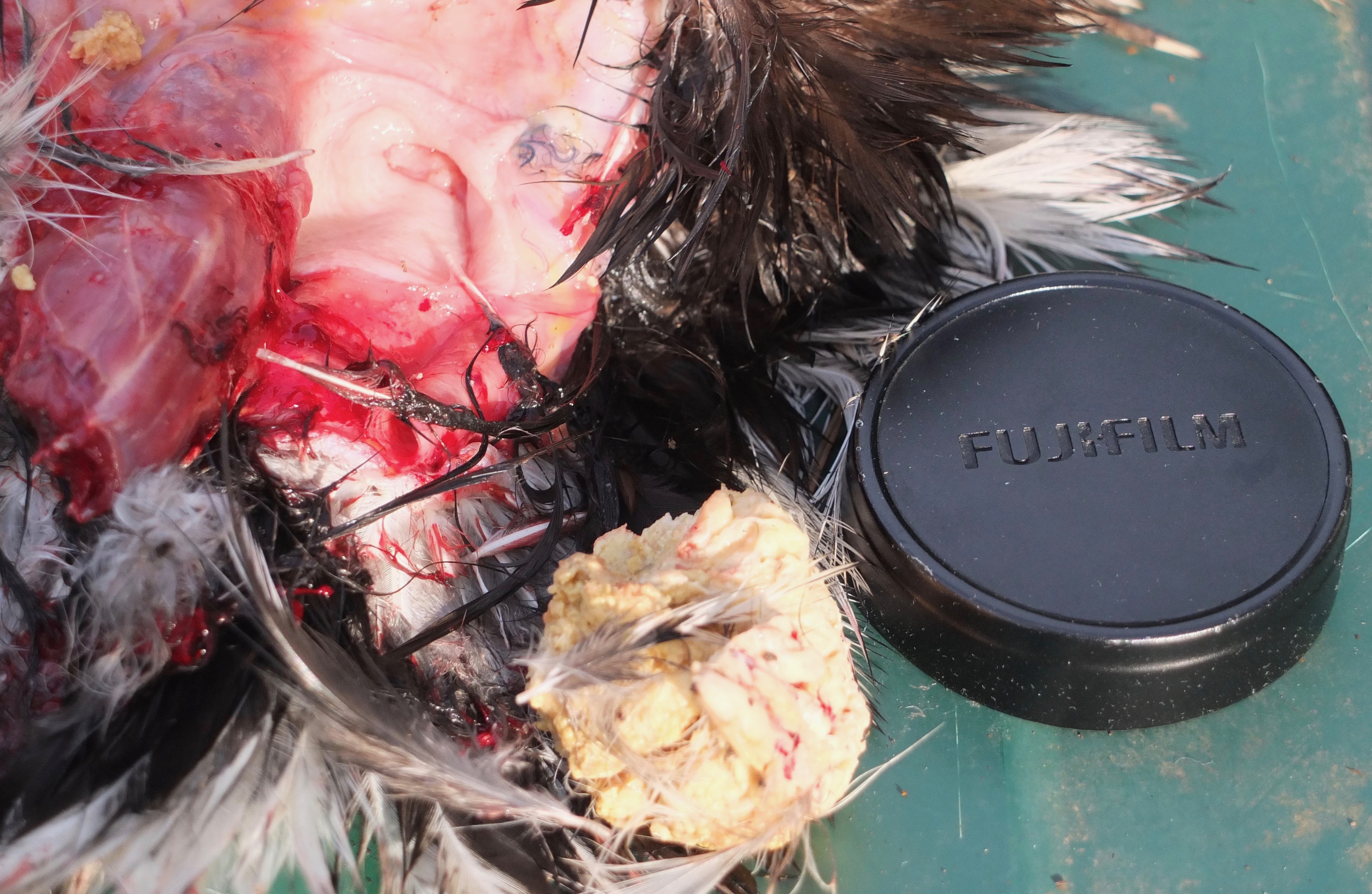
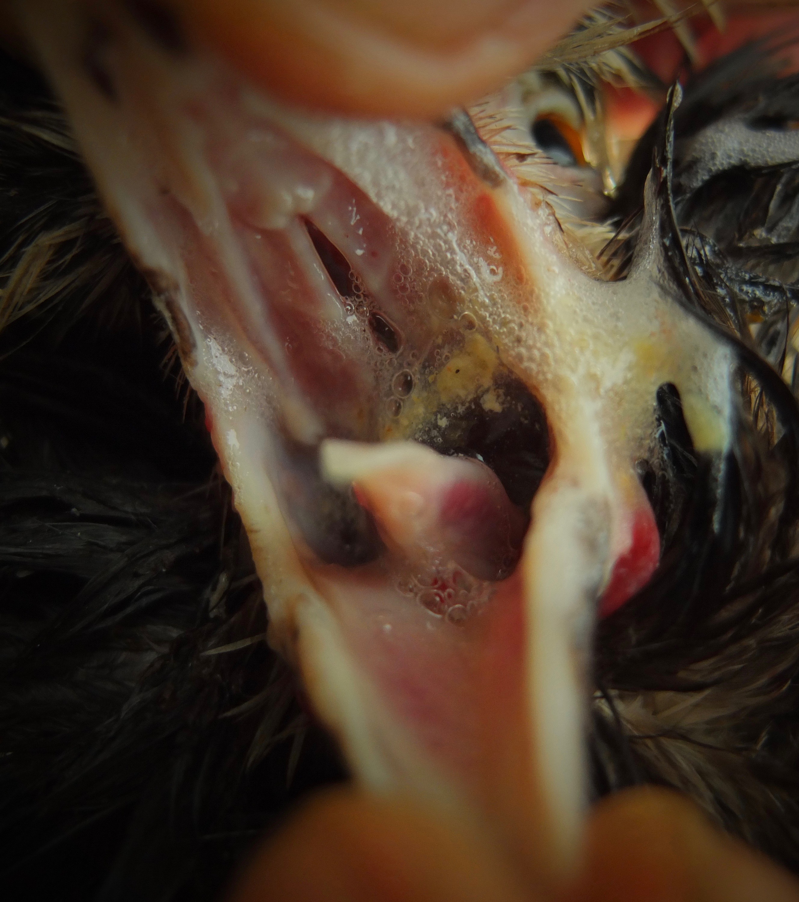
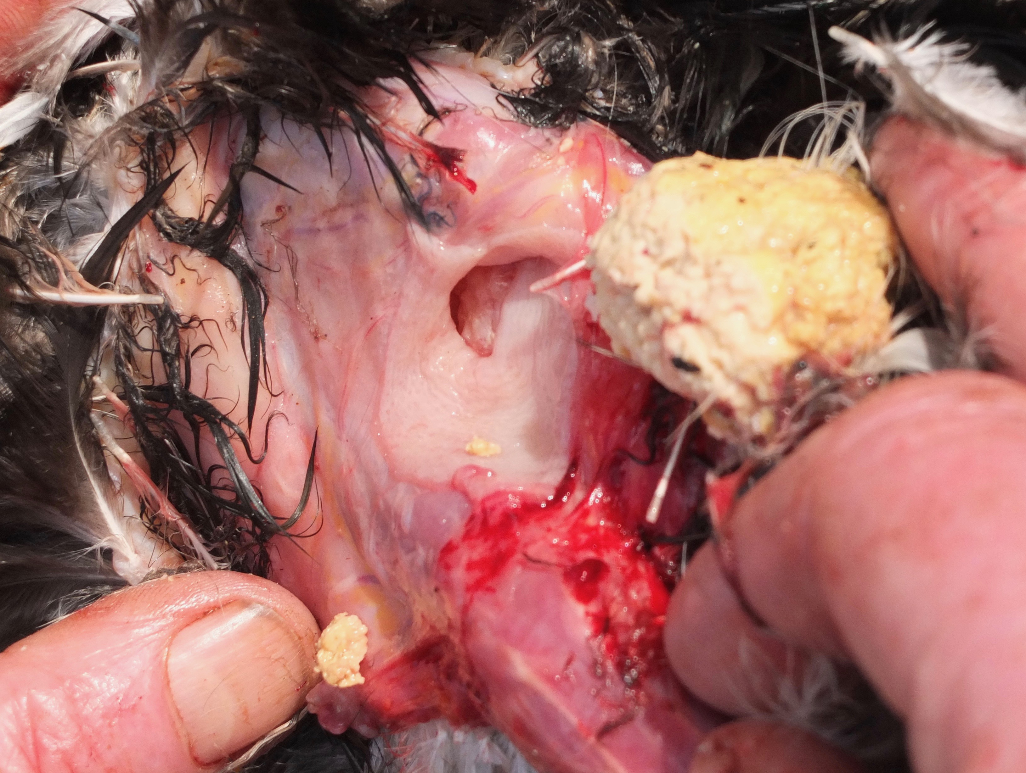
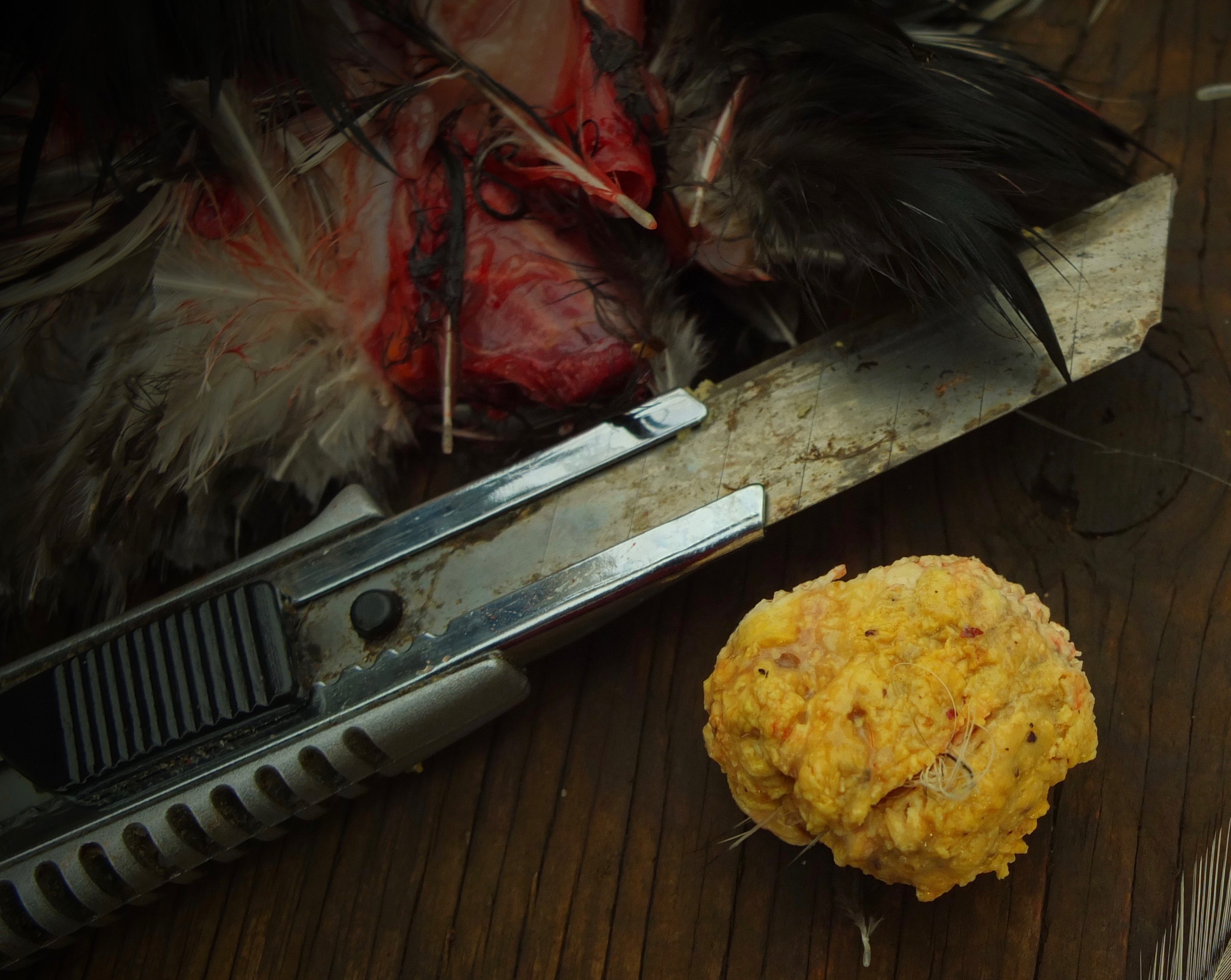

Dr Bowes: This is a trichomonosis infection caused by a protozoa, a normal inhabitant of the mouth. It can enter through breaks in the tissue from a penetrating injury or a scratch. It can be spread through dirty water. There is treatment but in this case it would have required surgery to remove the mass.
Glossary
Fibrin: an insoluble protein formed from fibrinogen during the clotting of blood. It forms a fibrous mesh that impedes the flow of blood.
Melanosis: a condition of abnormal or excessive production of melanin in the skin or other tissue.
Peritonitis: inflammation of the peritoneum, the lining of the abdomen
Serous: the clear liquid part of blood
Well, that wraps up another edition of Show & Tell With Bitchin’ Chickens and Dr Bowes. I hope that it’s been a learning experience for you.
If you’d like help with a case drop me a line using the ‘contact’ button on my home page. Remember to wear gloves, take good close up photos from several angles and supply us with plenty of information (e.g. timelines, symptoms, medications, general flock health, etc) so we’re able to more accurately pinpoint what’s going on.
Thanks again to Dr Vicki Bowes for her willingness to share her wealth of knowledge and experience to build capacity and skills in small flock keepers.
Featured photo credit: Stockai

Very interesting. Love it, learning so much. Thank you
LikeLiked by 1 person
More great cases!
LikeLiked by 1 person
Damn salpingitis! Hope it doesn’t hit another bird in my flock. Fingers crossed, everybody is doing well right now – Alicia
LikeLiked by 1 person