For more than three years I’ve gotten together with Dr Vicki Bowes, vet/ avian pathologist on a regular basis to pore over files in my memory stick loaded with interesting chicken health issues that I’ve collected for her expert opinion. She refers to it as ‘Show and Tell’, ‘Best Guess’ or, more recently, ‘Gorefest’ and has done a good job at making diagnoses given the information we have at hand. Sometimes all we are provided with is a short paragraph from the chicken’s owner, other times nothing more than a photograph.
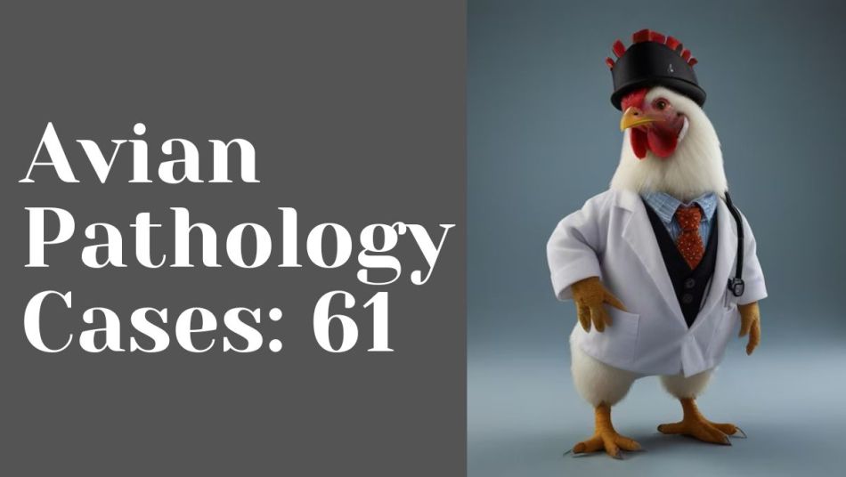
Herniated Oviduct
I thought my girl had an abscess on her thigh so I got my surgery tools out: clean x-acto knife, Vetericyn spray and Neosporin, and cut a small incision on the lower part of the abscess. It drained a little. I cleaned it and put her in a separate cage so she could recuperate. I woke up the next day to this! It seemed to fall out. She’s eating a lot and the feathers around the area continue to be wet, as if it’s still draining. – Leyla Keefe
Dr Bowes: The bulge could have been an abdominal hernia. To determine that you can push it in with your finger and see if it goes in or not. A hernia will go back in, but only temporarily. I think when you made the incision it allowed an opening big enough for the oviduct to herniate externally. The fiery redness is cause for alarm. It’s not black so it’s not necrotic yet. This would require a vet to stitch up the wound and treat with antibiotics.
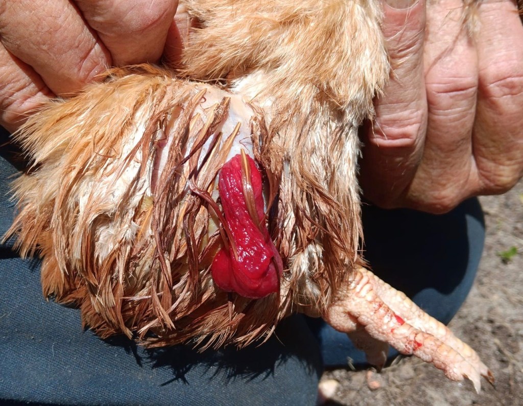
Bloody Egg
One of our 14 month old Leghorns was acting a bit off over the past 24 hours: close but slightly separate from the flock, eating fewer treats than usual, and often standing stiffly, as if hinged at the hips in a 90 degree angle, her head slightly tilted. She also had a streak of originally wet poop down her fluff, now dried and pulling at her feathers – not that uncommon for her, but this was black.
Yesterday afternoon, after I did some gentle water dabbing to loosen the dried poop, she immediately went to a nest box, and slept – unusual. She was one of the first to the roost, was asleep early, and didn’t open her eyes when I did the evening rounds.This morning I found this soft shelled egg filled with blood in one of the nest boxes, and assume it is hers. After this, she seems back to her usual self, with no new diarrhea. – Anonymous
As found in nest box (left) and cross-section (right)
Dr Bowes: This appears to be a hemorrhage off the ovary that has gotten into the oviduct. The blood is clotted and contained. The egg yolk follicle is encased in a vascular sheath with a weak point. As the follicle gets bigger it can cause a hemorrhage. It was probably just a one off but you should monitor her for any signs of pain or abnormal posture.
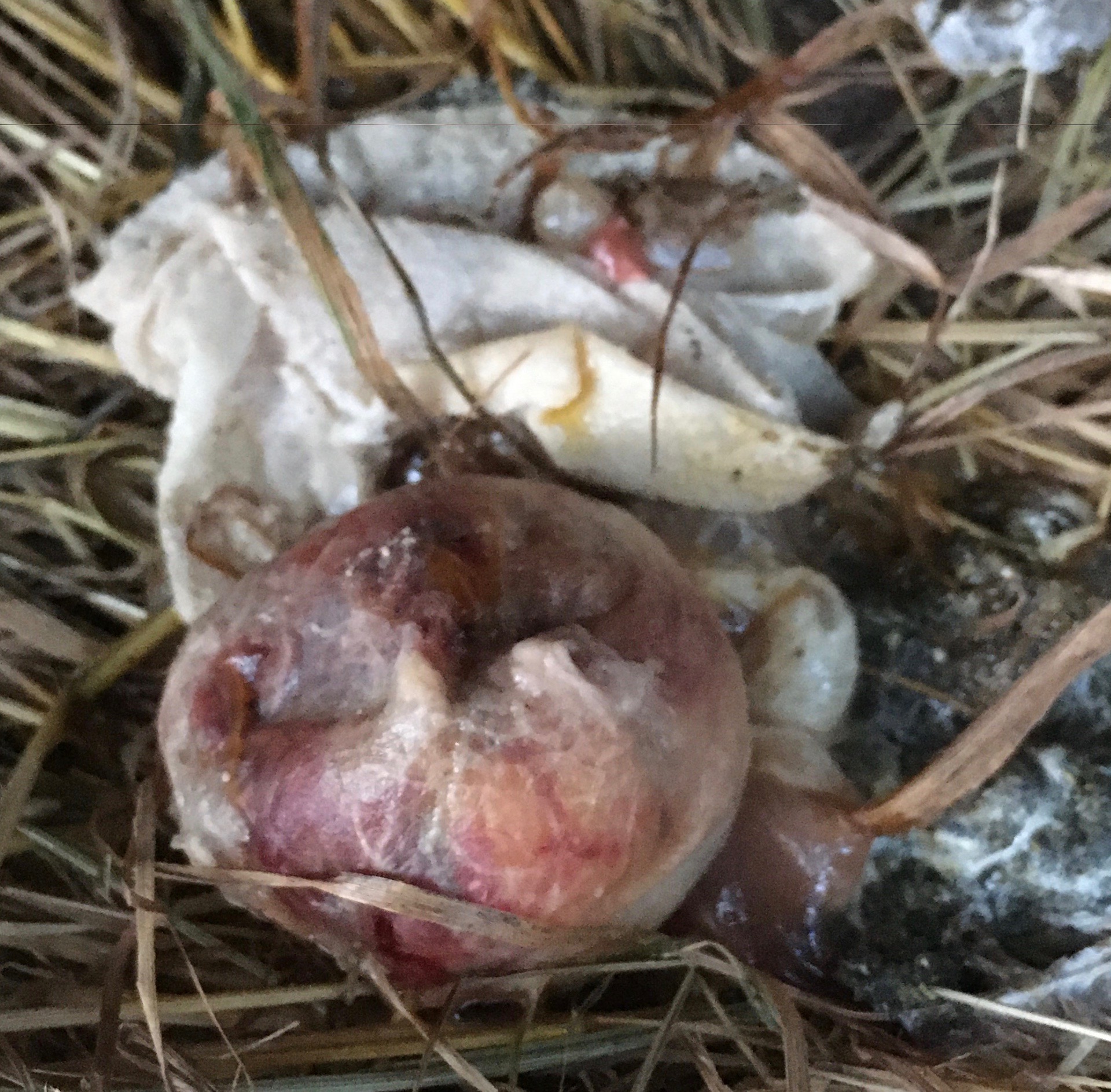
Mystery Object
I found this weird thing in the chickens’ wading pan. What is it? – Vincent Riggs
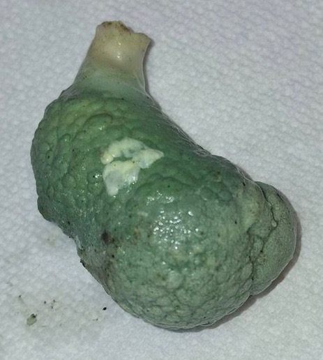
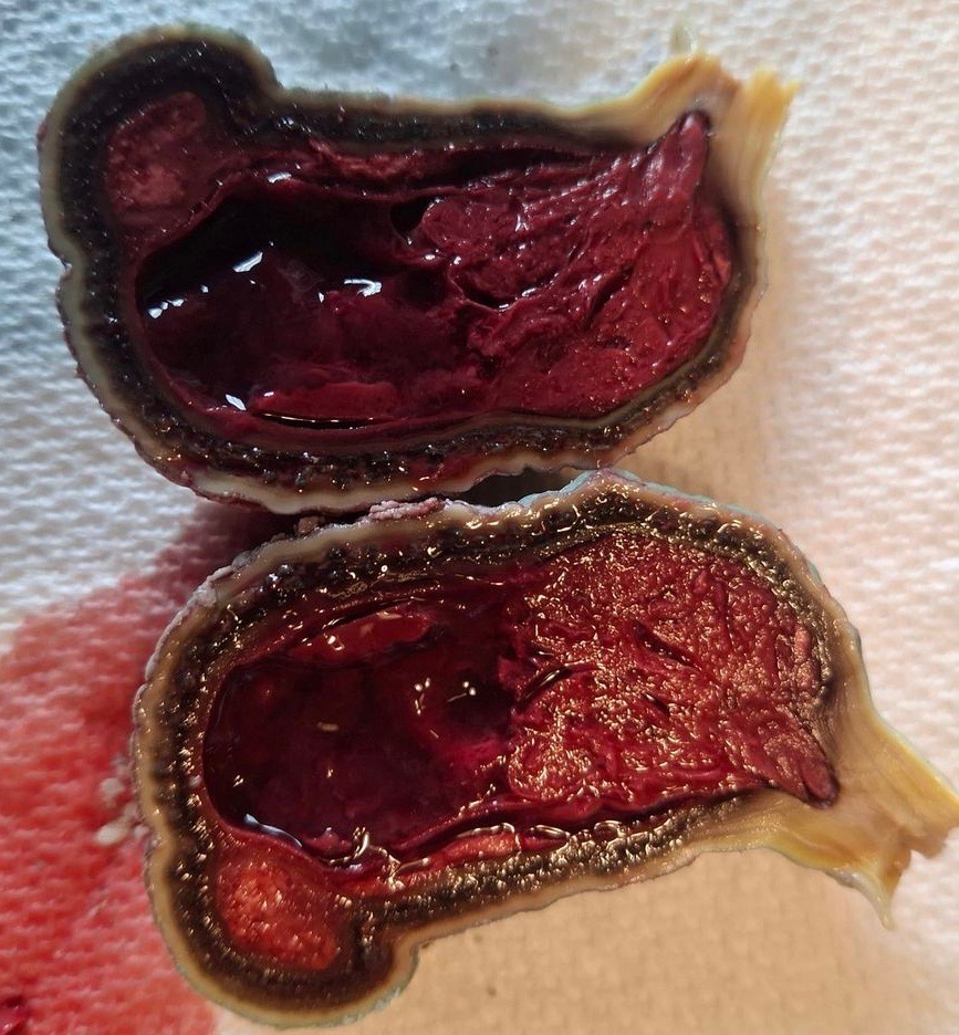
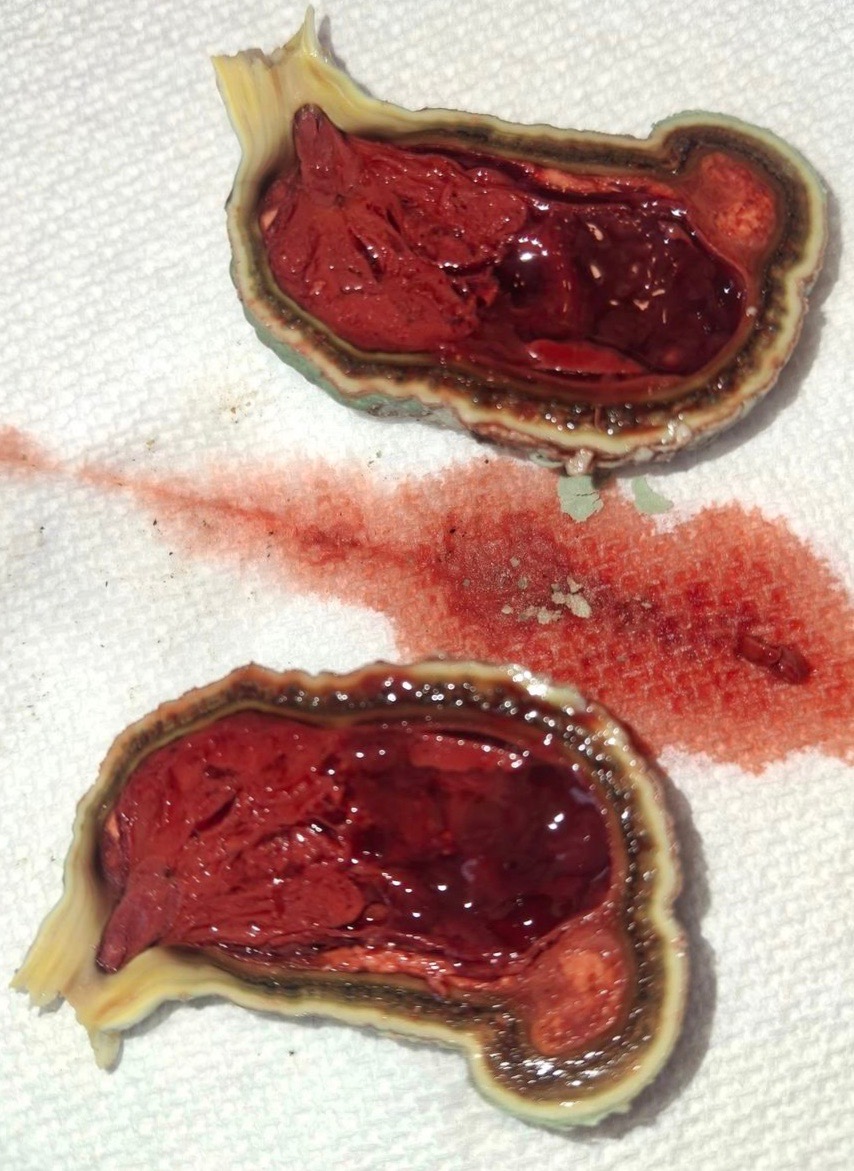
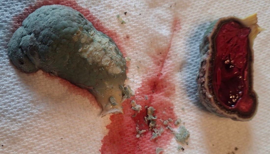
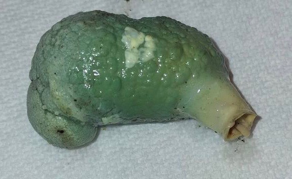
Dr Bowes: First of all, did this originate from your chicken? We’ll assume it did. I have no explanation for the interesting colour. The tubular structure and stem appear to be a cast probably from the cecal pouch and then been expelled. The layers around the outside indicate that it took some weeks to form and encapsulate the blood clot. Histology tests would indicate if there was a fungal or fibrin component to it.
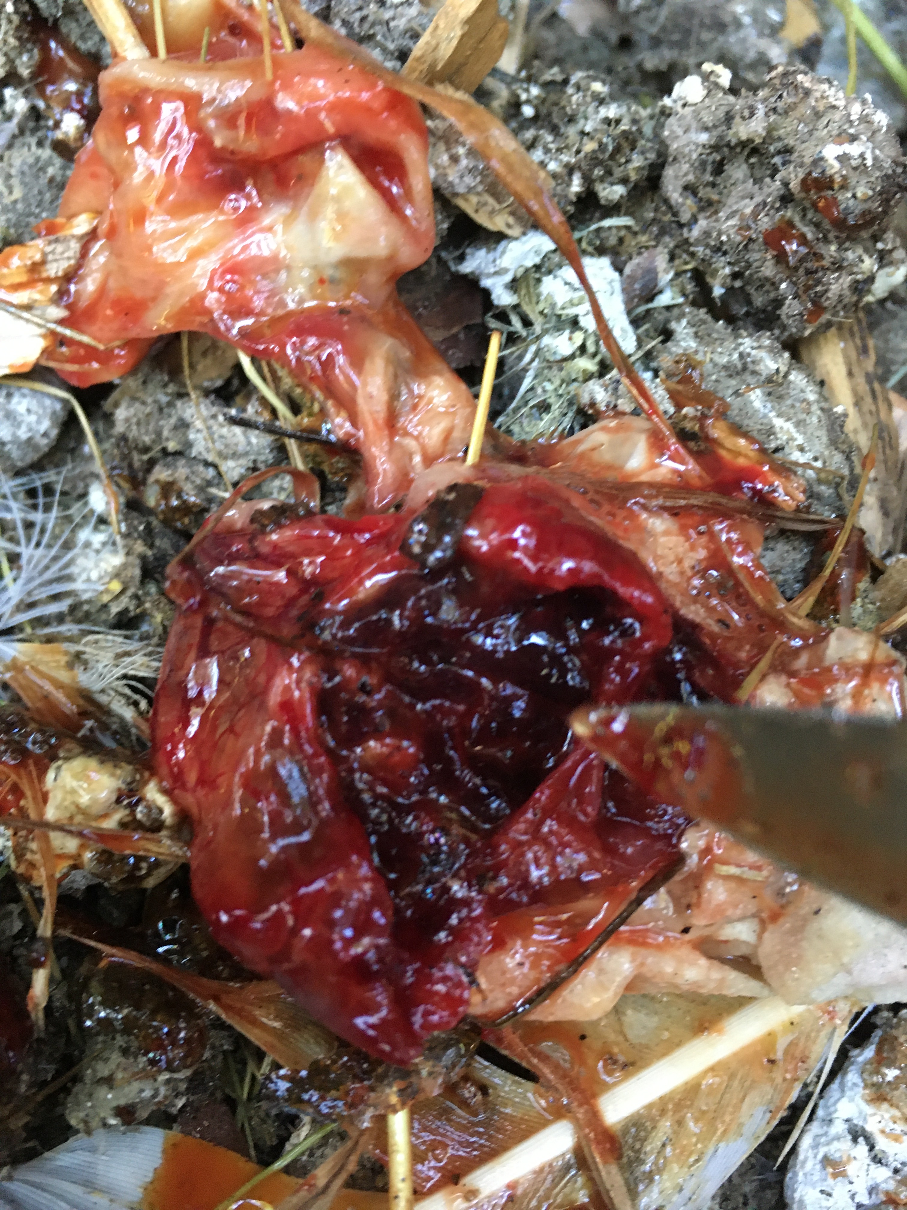
Salpingitis?
I find this on the coop floor. This blob was surrounded by egg yolk. I thought I’d be running a fecal float test on what I thought was poop in the center, but found it was very rubbery and when I pierced it with a pipette, some blood came out. It wasn’t easy to pierce because of its hard rubbery covering, the consistency of a gizzard. – Deb Watt
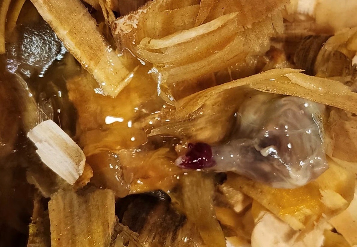


Dr Bowes: The ‘blob’ appears gelatinous with a tubular structure. Sometime during development an egg gets stuck and then continues on the process. There are yolk membranes and hemorrhage from the oviduct. It’s interesting that the components of the egg and yolk should be on the outside, but are on the inside. This might be, but is not definitive for, salpingitis.
Yolk Peritonitis?
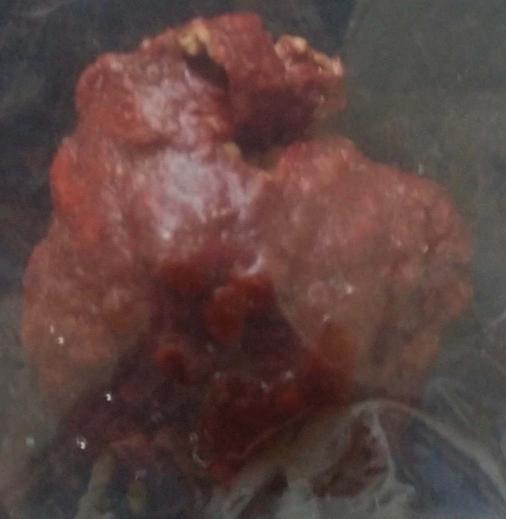
You’ll note that in some of the x-rays you can see a mass that looks like a ball of crumpled aluminum foil in her oviduct region. Those images are from the first visit, the ones with it missing are after she passed that obstruction. The mass itself is what’s in the Ziploc bag, both intact and after the vet broke it apart to examine contents. I wish we could have gotten a cross-section image, but at the time I felt it might be important for the vet to see exactly how it came out and cross reference to confirm nothing appeared to have been left behind.
Can you spot something important that our vet overlooked? We still haven’t been able to get a very clear picture of what’s going on apart from that blockage, but suspect something is, just based on the continuing digestive issues and apparent fluid still present at the second x-ray. I feel like she’s laying internally periodically and reabsorbing fluid between. Outwardly, she is acting like her system is on and functioning, but nothing comes of her efforts, so that has me concerned that she is doing something and it’s just not making it’s way out. I imagine septic EYP can be ruled out, as I don’t think we would be seeing these rebounds of full energy/appetite if anything had gone septic. She has good and bad days, and the bad ones always go hand-in-hand with the watery poop. She’s definitely got us scratching our heads.
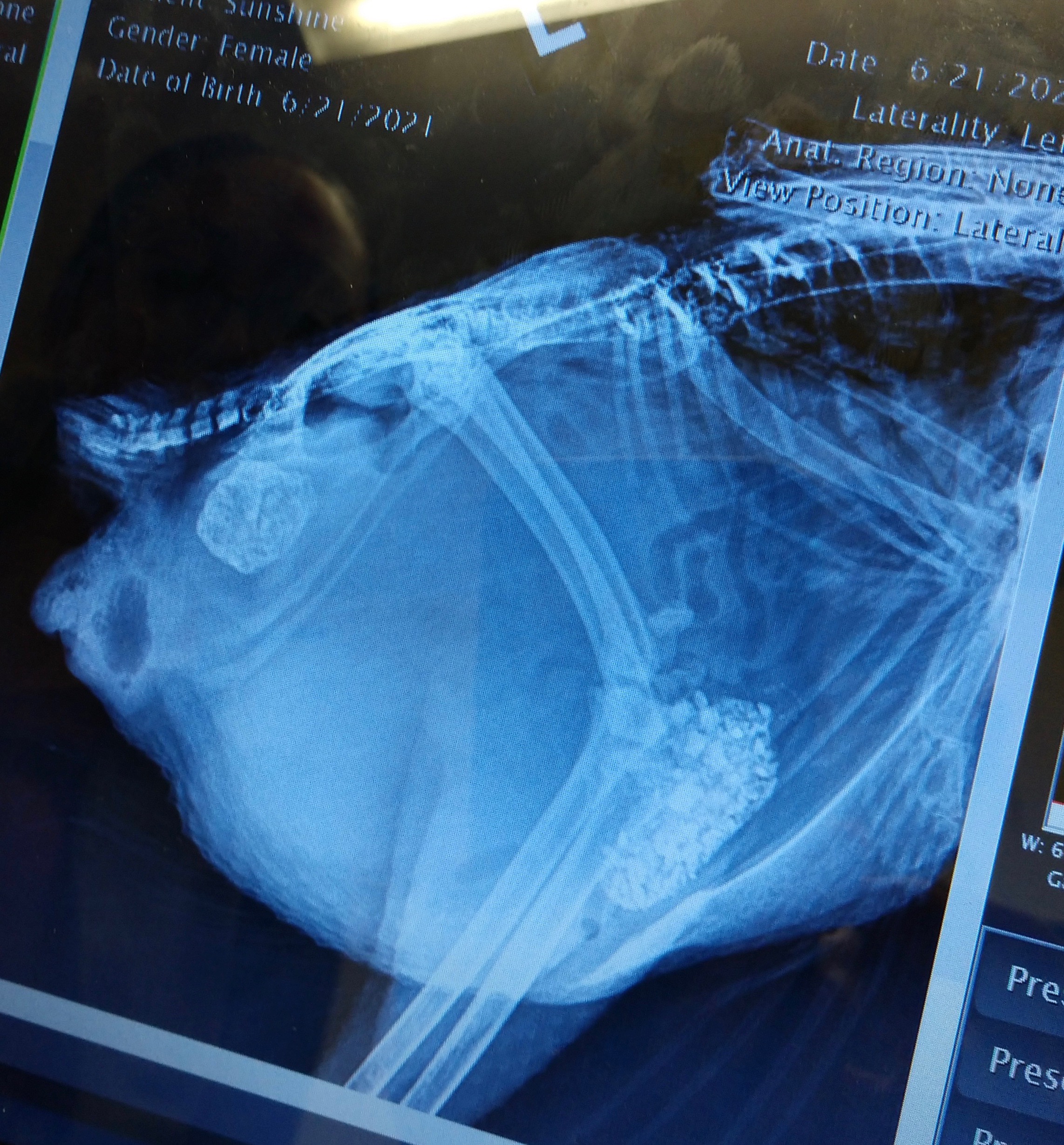
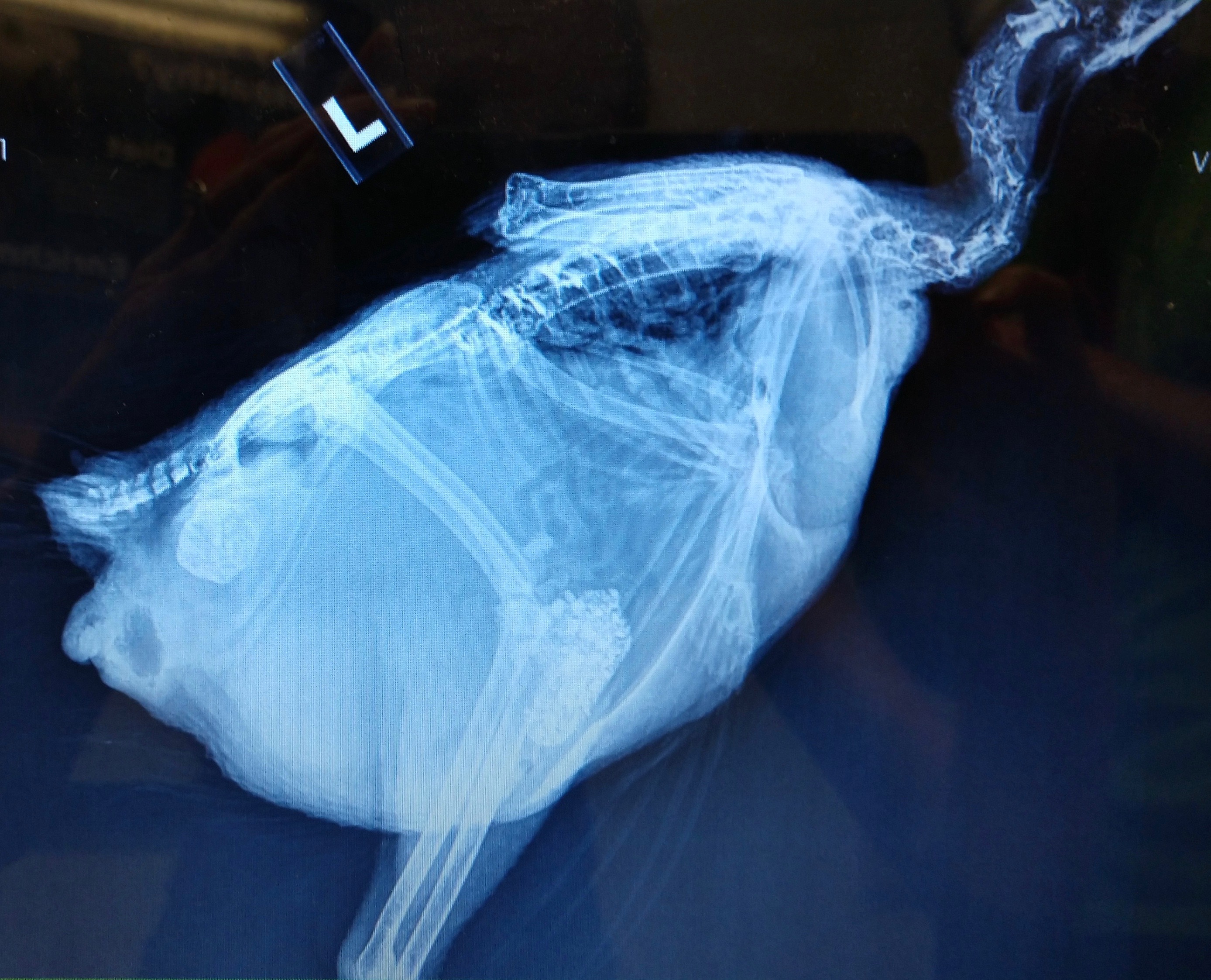
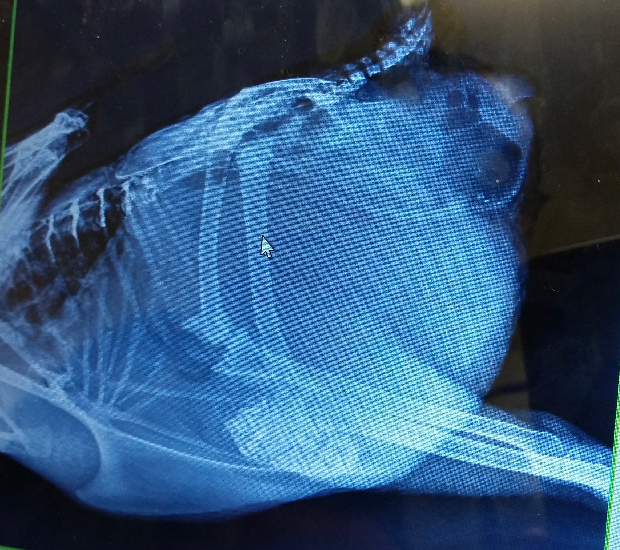
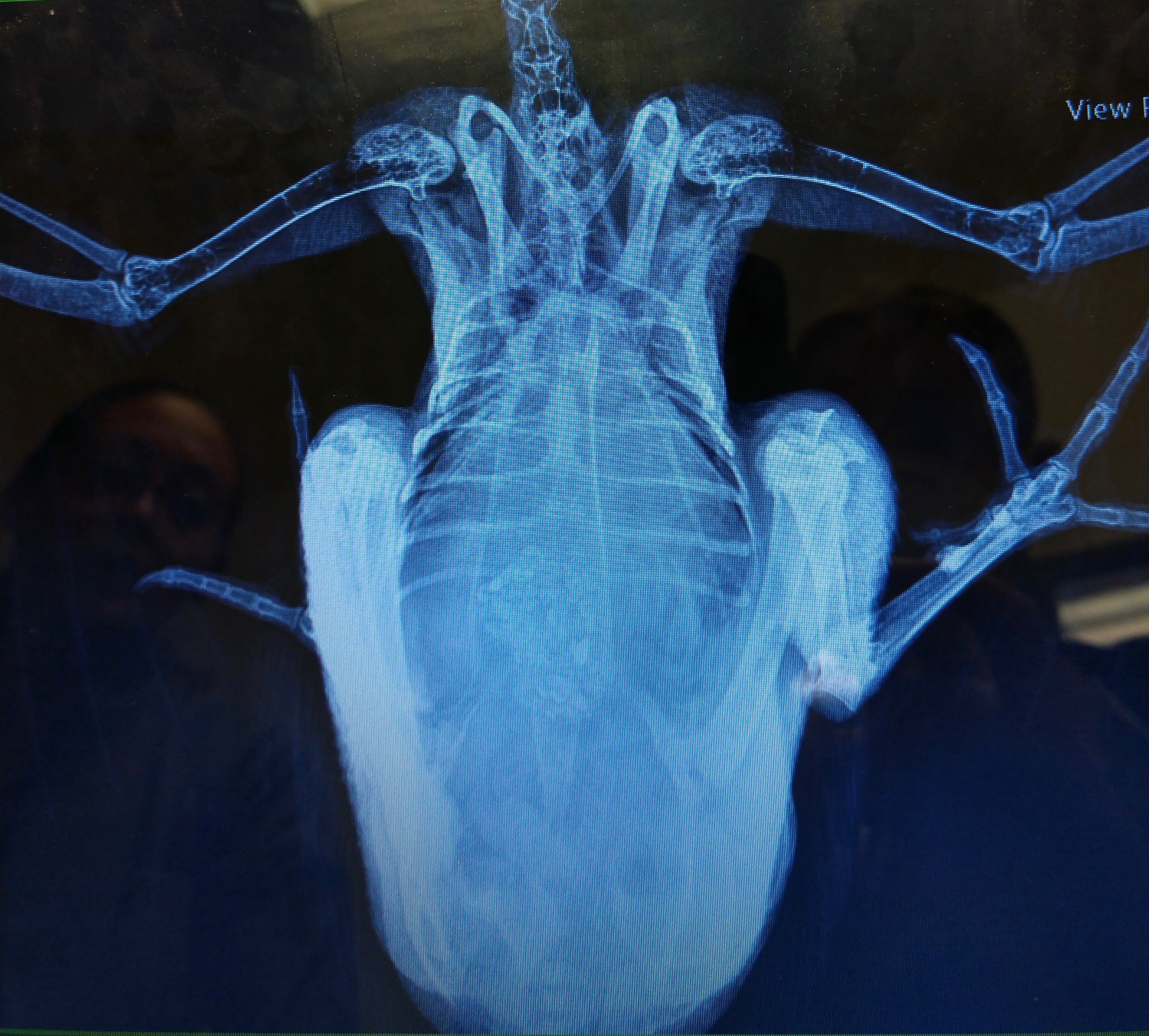

Dr Bowes: The vet did a great job of the x-rays. They indicate the ureters are full of crates, a sign of dehydration, the intestines look normal, there is a lot of internal fat. The mass is a collection of debris from the oviduct. It is crumbly and friable, the result of a hemorrhagic component. The intestines appear to be bunched up. There is something obstructing the full view of the abdomen. Is that fat or yolk peritonitis?
Foreign Body Ingestion
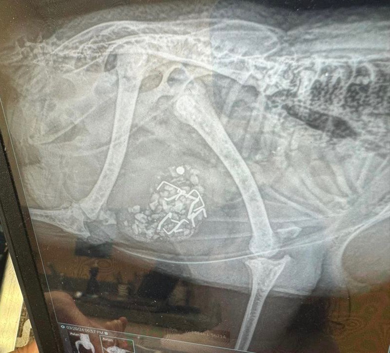
I noticed one hen laying down and not getting up. I found what I thought was broken yolk on her feathers around her bottom. I ultimately assumed an egg broke in her which I’ve dealt with many times. I scheduled emergency vet appointment with our avian vet for 6 pm. By then her leg is splayed out to the side, she’s deteriorated and seems more uncomfortable and won’t stand. They do X-rays and vet comes back in and says she has good and bad news (which should have been good and worse news). She didn’t have an egg break in her but looks to have eaten construction staples. As vet is showing me X-rays, tech comes in and says the leg or hip is confirmed dislocated. I decided to keep her hospitalized until Friday (when vet that could do surgery returned) for pain management and she received CaEDTA to absorb the heavy metals, next daywas eating and drinking on her own, and seemed to be perking up. Today she underwent a procedure to try to remove the staples using a scope with some robotic attachment, but they only were able to remove 1 of 8 without risking damage to stomach walls because they were embedded. They also popped leg/hip back in.
I’m picking her up tomorrow and she’s coming home on lactulose to see if it will help her pass the staples and back in 3-4 weeks for X-rays and another procedure/surgery with the scope to try to remove. I’m not understanding how if embedded they’d come out, going to ask more tomorrow when I get her, but moreso wondering if anyone has gone through anything similar. She was under anesthesia for 3 hrs, they said she did great but I’m also $2k in and I don’t want to keep putting her under, nor do I want to keep spending $1-2k to try to remove them. – Sarah Minderlein

Dr Bowes: I’m glad you took her to a vet but, unfortunately, I agree with your feeling that embedded staples will not come out on their own. Sometimes small screws or other objects can pass on their own, but these staples are fine with two sharp ends that will easily become lodged and not move down her digestive tract. Surgery is required to extract them.
Glossary
Histology: the study of microscopic structures of tissues
Well, that wraps up another edition of Show & Tell With Bitchin’ Chickens and Dr Bowes. I hope that it’s been a learning experience for you.
If you’d like help with a case drop me a line using the ‘contact’ button on my home page. Remember to wear gloves, take good close up photos from several angles and supply us with plenty of information (e.g. timelines, symptoms, medications, general flock health, etc) so we’re able to more accurately pinpoint what’s going on.
Thanks again to Dr Vicki Bowes for her willingness to share her wealth of knowledge and experience to build capacity and skills in small flock keepers.
Featured photo credit: Stockai

Fabulous article. Fortunately, so far, I haven’t had any of these issues. But, learned a ton. Thank you
LikeLiked by 1 person