This series is the result of my partnership with Dr Vicki Bowes, avian vet/pathologist. We met up four times in the fall to review some of the case notes I had collected for her opinion. These are some of the mysteries that she took a stab at solving.
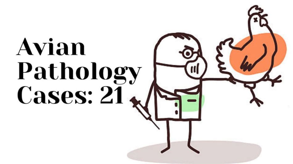
My 17-week-old Cream Legbar died suddenly with no signs of illness before I found her passed away.
She was a “runt” compared to the other pullet I got at the same time, but after she feathered up she seemed to catch up in size. However, once she had passed her breast muscle felt very atrophied. Her liver was swollen, very off color and appears like it has fatty deposits, protruding down into her abdomen. What is wrong with her liver? – Terri Smith Hubbard
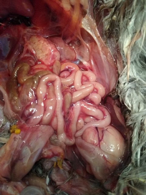

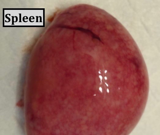
Dr Bowes: Her liver is massively enlarged and severely infiltrated with pale, tan tumours; the spleen is enlarged and pale; there are tumours in the ovary and kidneys. Diagnosis: Marek’s Disease.
Abnormal Structure
Someone gave us a chicken that has this growth on her neck. She seems healthy and eats fine. Is there cause for concern? We definitely don’t want her suffering. – Dan Tangen

Dr Bowes: I would be interested to feel that structure to confirm if it is made up entirely of skin or there is any involvement of bones. It is fully feathered and looks healthy. I would say this is an anomaly, perhaps caused by misplaced embryonic tissue, but doesn’t seem to be impeding her quality of life. Let her life out her life and enjoy your freak of nature.
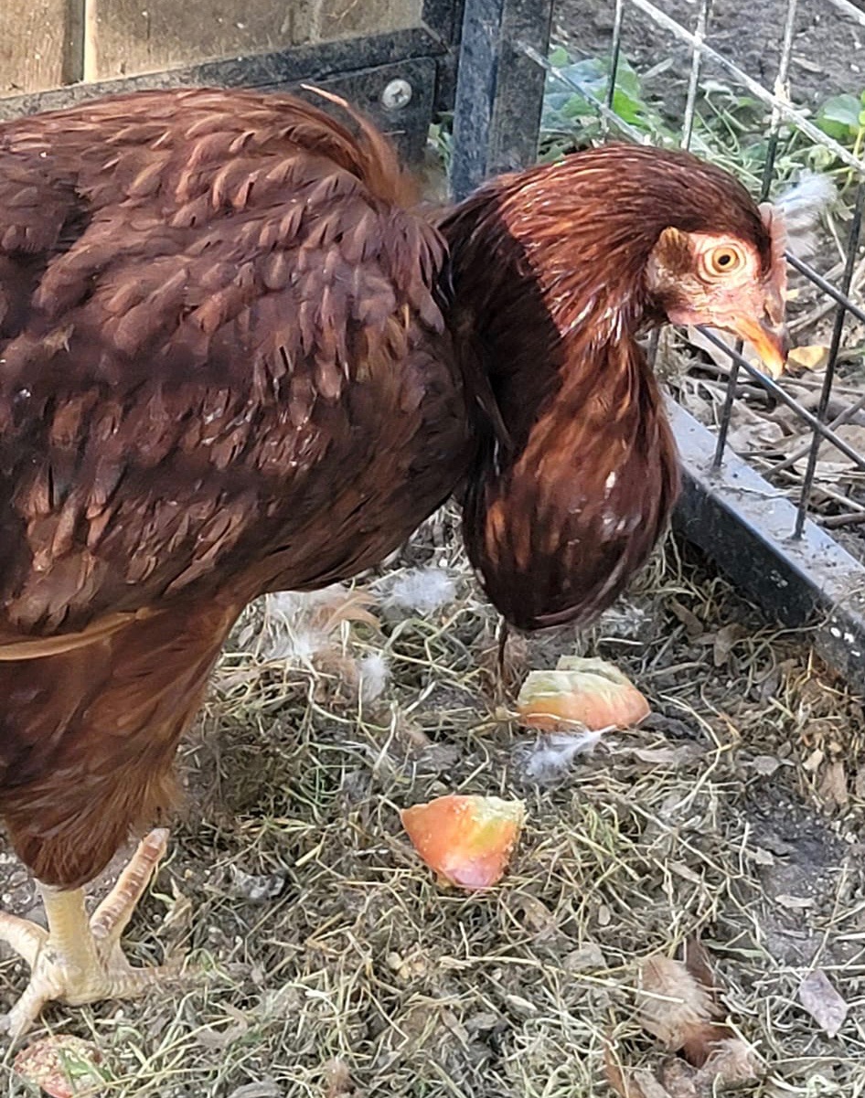
This just happened to my hen. What can I do? – Destiny Hambrock
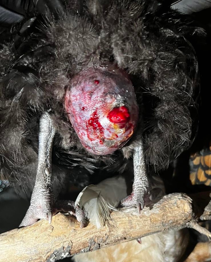
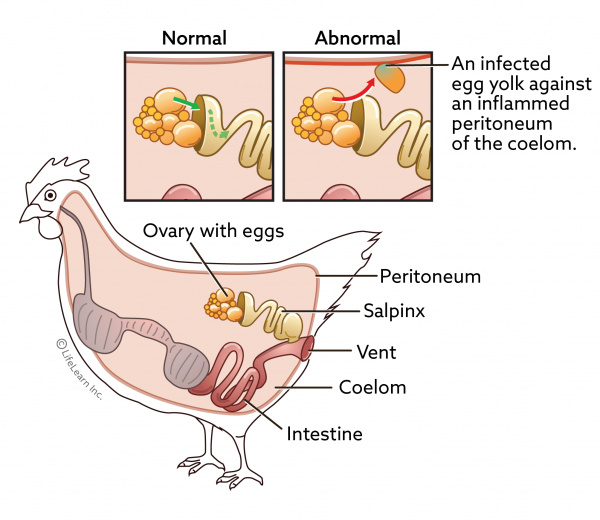
Dr Bowes: The abnormal swelling of the abdomen encourages other birds to peck at the area. I would diagnose peritonitis with the secondary issue being cannibalism. Recommendation: humane euthanasia.
What Is This?
“What could this be? I thought it was a chunk of mud on one of my hens beak and picked it off.” – Danielle Partin
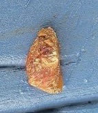
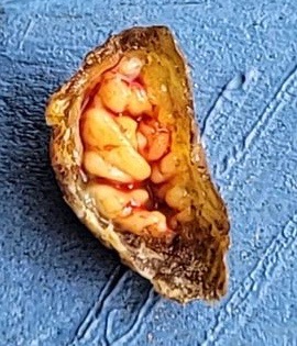
Dr Bowes: It looks fatty with normal components of skin and keratin. I’d like to see a photo of what the beak looked like after it was removed.
Tumours
One of my Columbian Wyandottes had water belly (ascites). As I was getting ready to drain it, she died. I decided to open her and check what was going on just so I could learn more. What are these things on my chicken’s small intestine? – Daiene Bagarini Ferguson
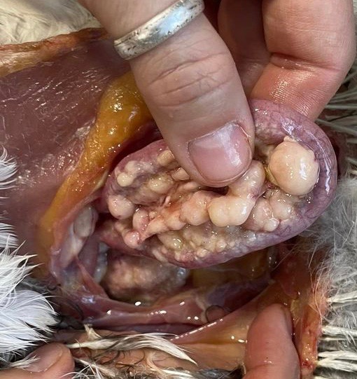
Dr Bowes: Those are caseous nodules in the mesentery of her intestines. You’d need to cut into it in order to diagnose what was going on. If it looked the same on the inside then it would be neoplasia (tumours); if it was caseous then I’d diagnose it as an abscess.
If there were nodules with a cheesy core in other filtering organs like the liver and spleen then I’d test for tuberculosis (TB).
Well that wraps up another edition of Show & Tell With Bitchin’ Chickens and Dr Bowes. I hope that it’s been a learning experience for you.
If you’d like help with a case drop me a line using the ‘contact’ button on my home page. Remember to wear gloves, take good close up photos from several angles and supply us with plenty of information (e.g. timelines, symptoms, medications, general flock health, etc) so we’re able to more accurately pinpoint what’s going on.
Thanks again to Dr Vicki Bowes for her willingness to share her wealth of knowledge and experience to build capacity and skills in small flock keepers.
Credit featured image: The New Yorker

0 comments on “Avian Pathology Cases: 21”