
This series is a partnership between Bitchin’ Chickens and Dr Vicki Bowes, vet/avian pathologist. We regularly get together to chat about interesting chicken health issues that I’ve collected and attempt to come up with a diagnosis based on the information we have, which often isn’t much.
Liver Tumour
This enlarged liver came from a two-year-old Welsummer hen, who presented with lethargy and a pale comb. The entire liver is affected: wrong color, texture, and is finely filled with infiltrates and small and large foci. – Lorri Dee
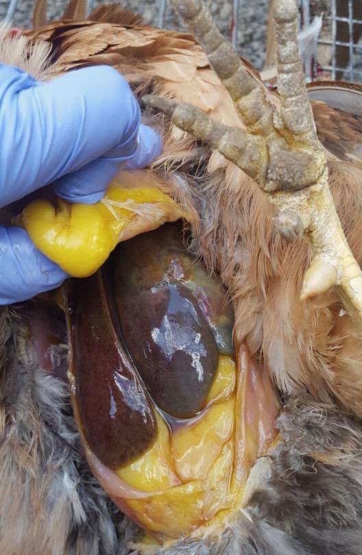

Dr Bowes: My first inclination is the liver was affected by a rapidly growing tumour. The margins of the organ are rounded, not sharp and there are areas of necrosis. I would have liked to have seen photos of the spleen. Microscopic work would be required for an accurate diagnosis.
Suspected Fungal Infection
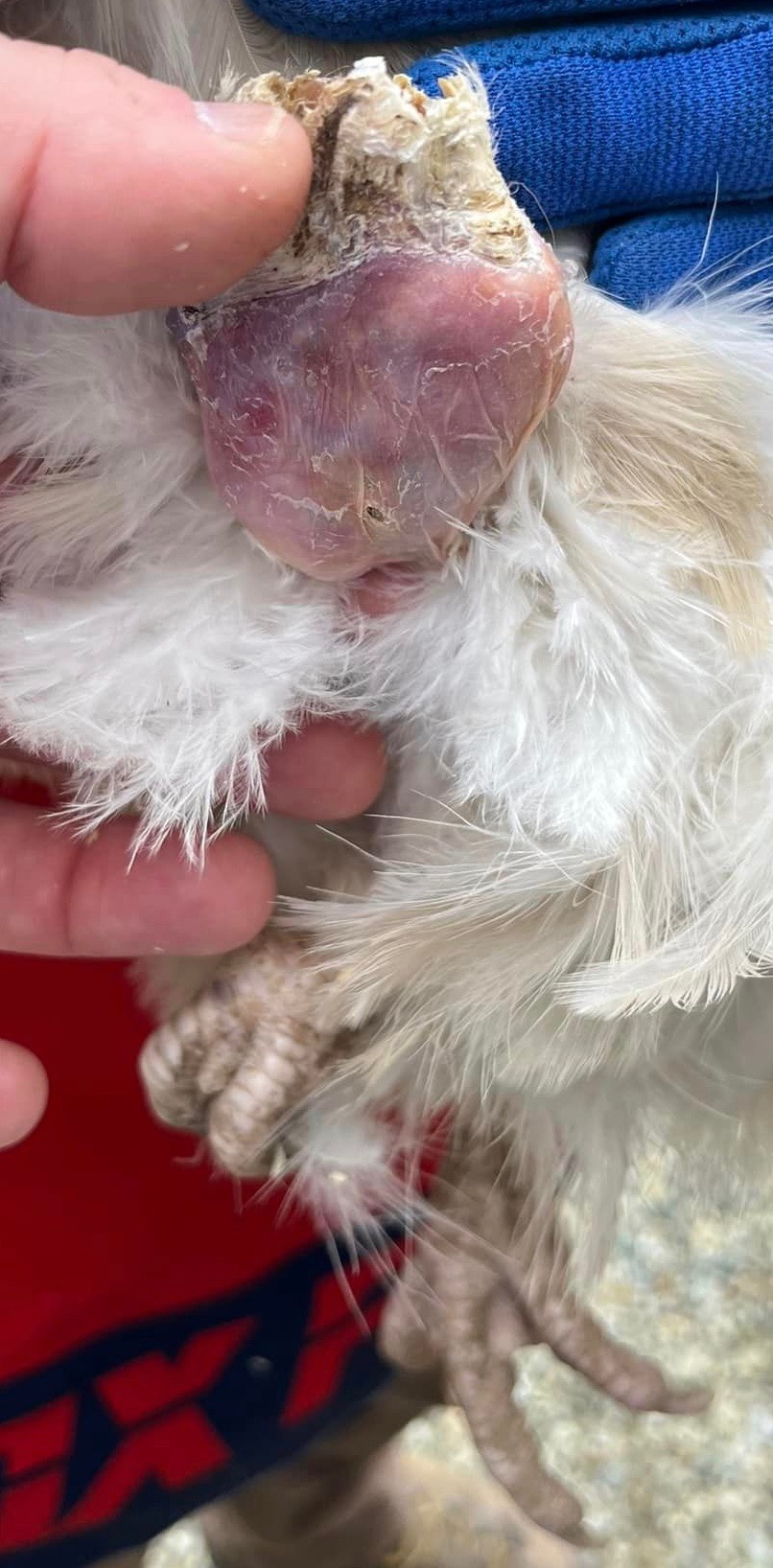
Our Silkie hen has an interesting skin/follicle issue. Our vet is stumped because skin cultures were negative for bacteria, fungus, and parasites/mites. It is not a bot fly in her ear, but it is very swollen. The yellow projections are like her feather follicles; they’re quite hard and a bit waxy. It started as a scab looking lump of tissue on the back of her neck when we adopted her this summer. She is in good spirits and acting relatively normal, but her condition has been getting worse. It doesn’t appear to be contagious to other chickens. – Marybeth Hopperton

Dr Bowes: I would like to know what cultures were done to rule out a fungal infection, which is what I would suspect. Remove all the waxy material manually and have a culture done of the ear infection, which is the priority so the infection doesn’t enter the brain.
Feather Inclusion Cyst
I found this growth hanging down by my Orpington’s foot, which I didn’t notice because she’s so fluffy. I gave her an Epsom salt bath and was able to open it up and pulled out a bunch of what looks like debris, which was very stinky. No bleeding. Soaked it with antibacterial solution and she is now resting. – Kylee Atherton Irg
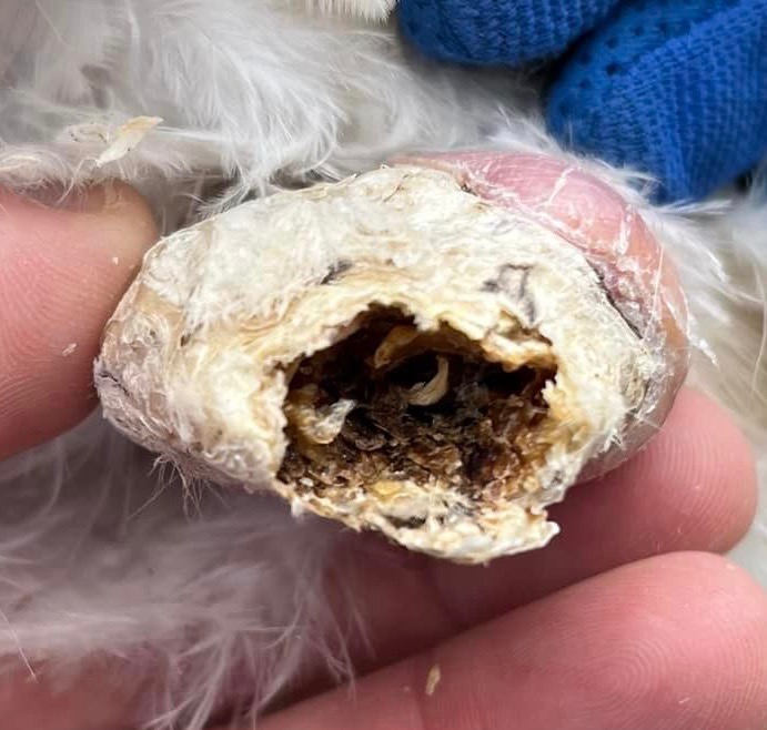


Dr Bowes: This looks like a slow growing plug called a keratoacanthoma or feather inclusion cyst. It is a vascular structure so I recommend veterinary care to remove the whole lump; if not, it may regrow. Pain management and antibiotics are required.
Sinus Infection
I do rescue and rehab in Hawaii and I had a young cockerel that had a severe sinus infection. For two months I couldn’t treat it, even with the strongest of prescription meds.The lancing was done twice by a vet who has no experience with birds. As we know, this kind of infection turns to hard pus quickly as it makes its way to the only exit, which is typically the eye socket.
Since there were now open incisions the infection stayed in the cheek cavity and festered. He was never without some sort of tetracycline antibiotic or a broad spectrum such as Baytril, but nothing helped. I even tried to flush with Tylan wash without success. I don’t know how this poor thing just acted normal everyday. I finally did a mercy cull to see why medications wouldn’t reduce the amount of infection and swelling and then opened the cavity. I’d most like to know if I could have injected Tylan or a Tetracycline directly into the face. – Cynthia Higa

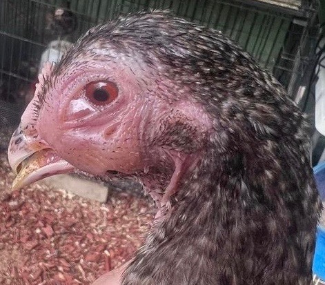


Dr Bowes: This was treated as sinusitis, which appears that once it was resolved became cellulitis. The vet didn’t make things worse by lancing the area – that is the only way to remove the kernel of infection.
It would have been useful to have done a culture to determine what kind of infection he had. Baytril is an effective antibiotic, but not good for Staphylococcus. If that was the case Penicillin would have been better. Injectible medications wouldn’t have been more effective.
Cystic Oviduct
My hen was acting sick and lethargic. I gave her a bath to soak her vent to clear some pasty butt and this came out several inches below her vent leaving a round hole. What caused this? I assume the skin was necrotic/there was an abscess. The mass is very large, bigger than my hand with my fingers spread open. – Aimee Jones
Bitchin’ Chickens: I came across this case in an online chicken group and was pretty shocked at what appeared to ‘drop’ out of the poster’s hen. It resembled a breast implant which Dr Bowes referred to it as a ‘space-occupying lesion’ (SOL).
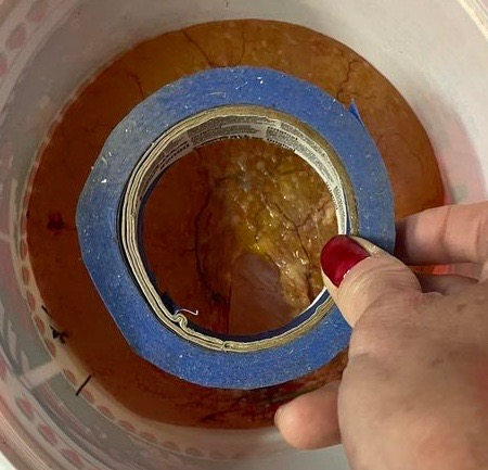
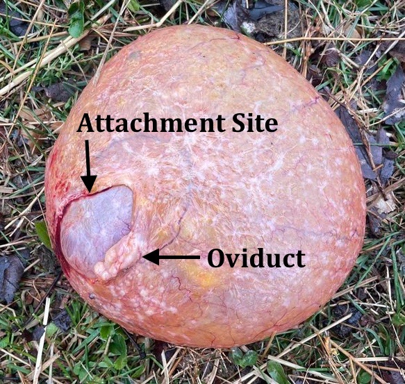

Dr Bowes: This appears to be a slow growing cyst, which may have taken months to achieve this size. The light area look like an oviduct and the dark spot may have been attached to the abdominal wall. It’s a fibrous capsule with well-formed vascularization. I am curious if it was attached to the outside of the oviduct and when she last laid an egg. Infectious Bronchitis can be linked to cystic oviduct.
Bitchin’ Chickens: I contacted Aimee for more information. She doesn’t know when her hen last laid, or if she ever did. She said she noticed that the tops of her hen’s legs were very large and once the cyst was out, not surprisingly, they returned to normal. Aimee sent me a video of her cutting open the fluid filled mass – it looked relatively clear with nothing of note coming out of it. Unfortunately her hen died a few hours afterwards.
Glossary
Cellulitis: deep infection of the skin caused by bacteria
Fibrous: tissue that is mostly made up of tough protein fibers called collagen and cells called fibroblasts.
Keratoacanthoma: crater-like ulcers with raised margins within feather tracts that usually contain a mixture of keratin, cell debris, and bacteria
Vascular: relating to, affecting, or consisting of a vessel or vessels, especially those which carry blood
Well that wraps up another edition of Show & Tell With Bitchin’ Chickens and Dr Bowes. I hope that it’s been a learning experience for you.
If you’d like help with a case drop me a line using the ‘contact’ button on my home page. Remember to wear gloves, take good close up photos from several angles and supply us with plenty of information (e.g. timelines, symptoms, medications, general flock health, etc) so we’re able to more accurately pinpoint what’s going on.
Thanks again to Dr Vicki Bowes for her willingness to share her wealth of knowledge and experience to build capacity and skills in small flock keepers.

0 comments on “Avian Pathology Cases: 25”