I met up with my mentor Dr Bowes, avian vet/pathologist twice in April. The first time we worked through dozens of cases for my series on avian pathology issues and the second time she performed necropsies on three of my hens that had died over a period of five months that I had stored in my freezer.
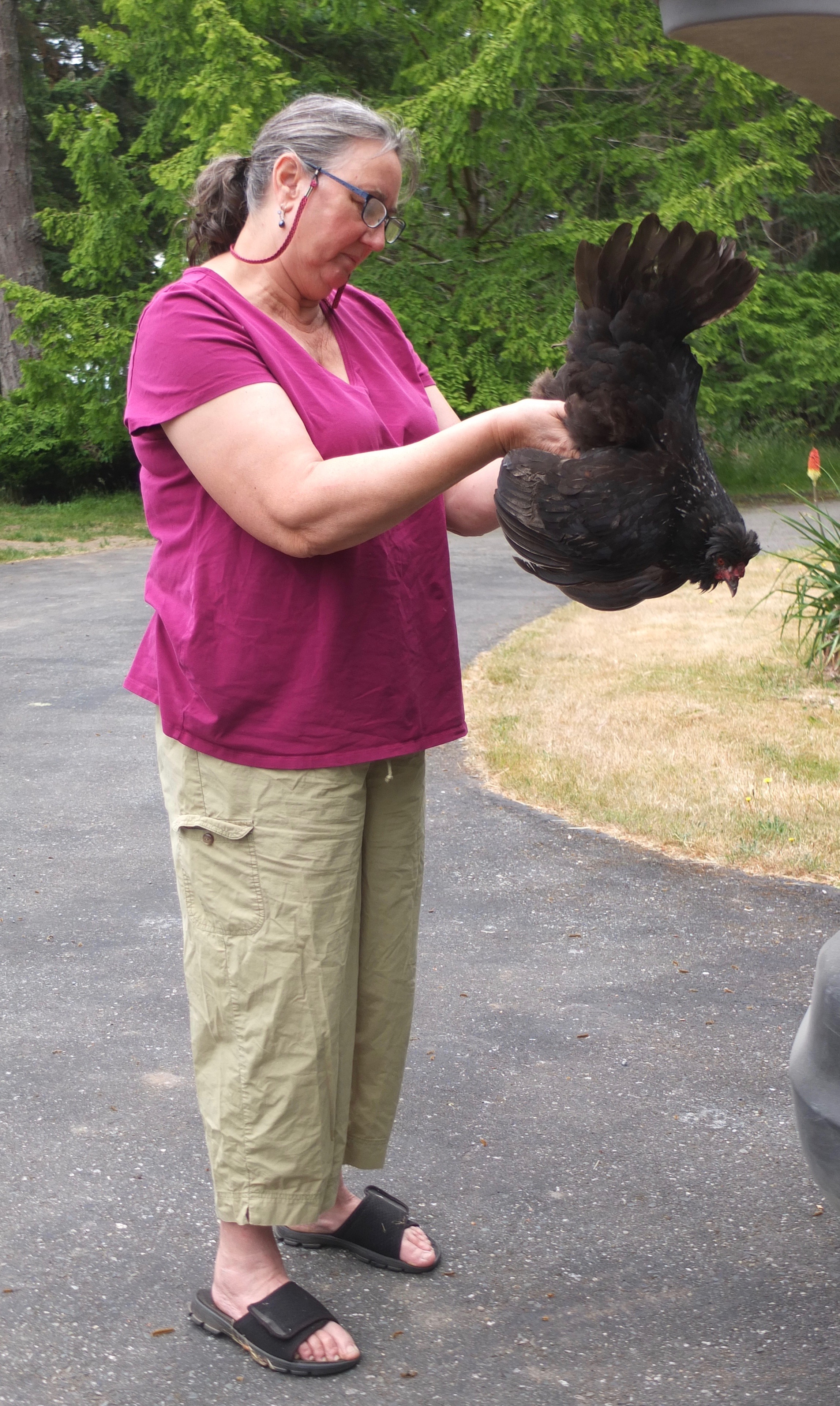
Last May was a bit of a unexpected combo: just before I was heading over to her place to work on new cases I went to check my flock and saw one of my hens standing in an awkward position that screamed ‘major illness’. She didn’t move as I approached and let me pick her up without a struggle. Her keel was prominent and I knew she was suffering from something that I wasn’t able to cure.
I left a quick voicemail for Dr Bowes explaining the situation and said I was heading over with my hen, assuming she was ‘a goner’. Upon our arrival she did an assessment which included looking at her eyes, in her mouth, at her skin, feeling her keel, and moving her about to see how she responded. The conclusion was she didn’t have the ocular form of Marek’s Disease, she was emaciated, non-responsive to normal stimuli and she had lice. The latter was surprising since I’ve rarely seen them on my birds. I assumed it was the result of her not dust bathing regularly like the rest of my flock.

She concurred that my hen was terminally ill, and asked how I wanted to proceed. I’m not unrealistic about a bird making a miraculous recovery so I asked if she would euthanize her and do a necropsy. Dr Bowes did so using the cervical dislocation method, which was very fast. She held her for several minutes pot-mortem as my hen flapped and jerked around, the result of electrical impulses and air leaving her body. When she was still a considerable amount of fluid leaked out of her beak, pooling around her.

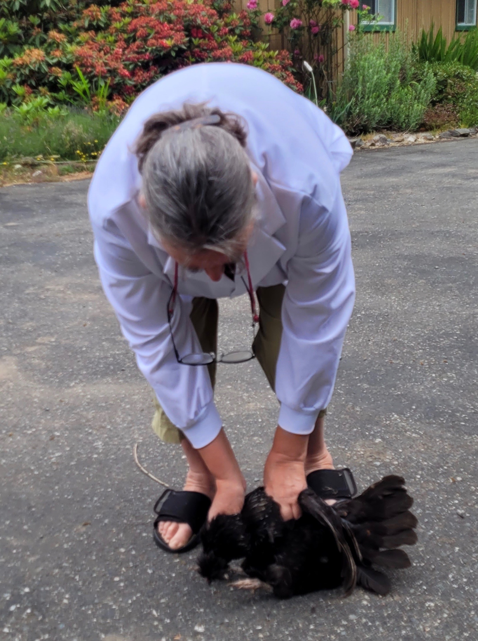
Six week before we had set up a makeshift dissection workstation on the tailgate of her truck and that’s what we did again. This time it was overcast and made for taking better photos.
Molly, Appenzeller Spitzhauben x Easter Egger, 3 years old
Some history: I have a flock of about 32 hens and one rooster. About half are under the age of two, while half are between three and six years old. Molly fell in the middle. In every flock some birds distinguish themselves by being friendly, or loud, or having unique plumage. Again, she fell somewhere in the middle. I think she laid a blue egg but I’m not sure.
Looking back on my records, it was about two months before when I noticed that she had accumulated poop around her vent. I cleaned it off and clipped some of her feathers in that area. I kept an eye on her, but didn’t notice anything unusual. She was still eating, drinking and roosting like normal right up to the end. If I had picked her up more often I would have felt that her keel was becoming more prominent and she was losing weight. There was nothing I could have done to treat her, but I could have euthanized her earlier.
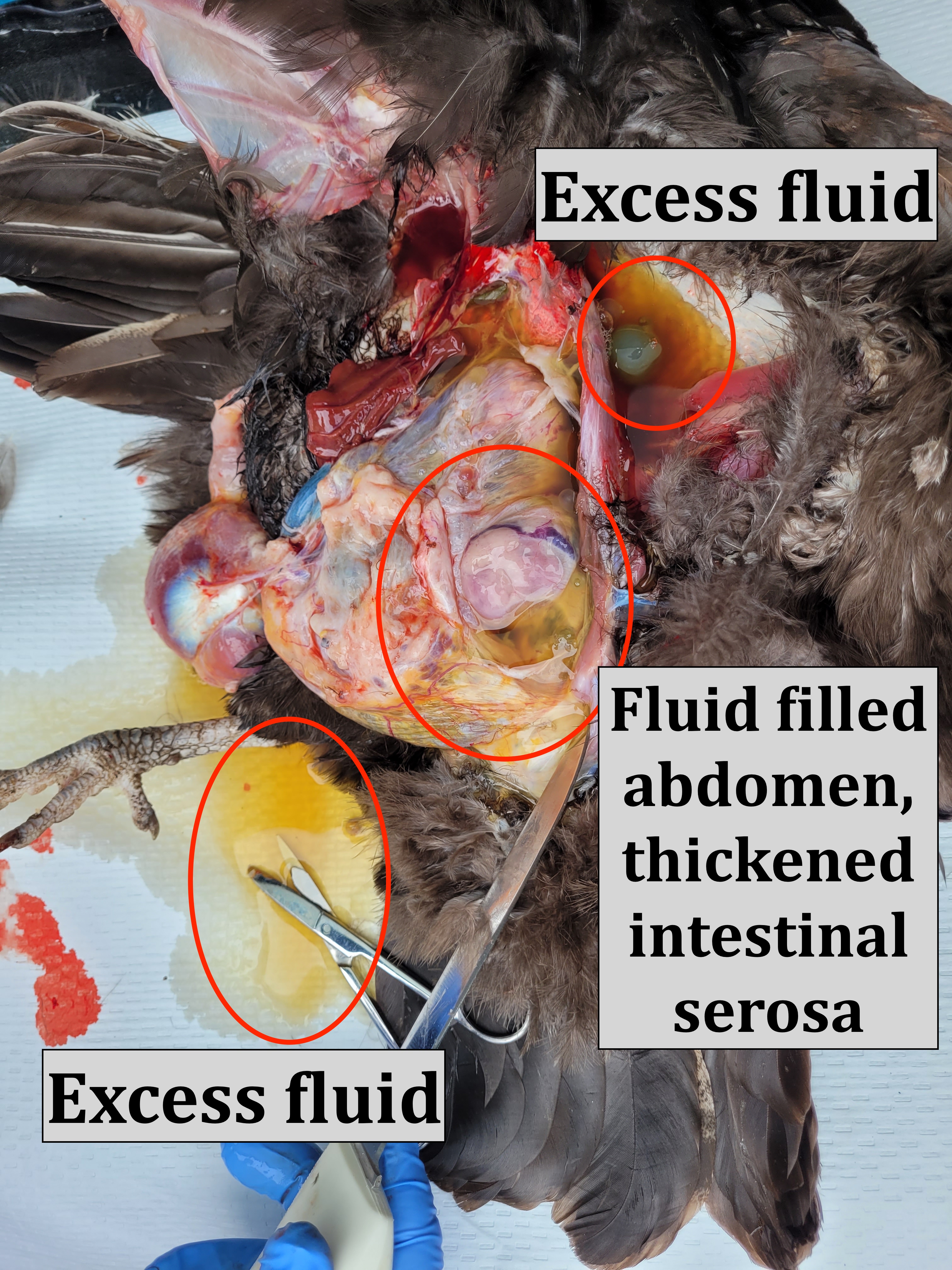
It’s been a great learning experience for me to watch someone who is skilled at what they do. Dr Bowes made incisions in the body to release the wings and the keel. What we were left with was a translucent membrane that held everything within the abdomen in place. She carefully cut the skin away but left that membrane intact as she predicted that fluid would leak out. She wasn’t wrong; a great gush of yellow liquid poured out once the membrane was pierced. It was similar to the fluid I’ve seen in cases of ascites (water belly), but Dr Bowes said the origin was different.
She carefully removed various organs, made comments about each of them and explained the significance of what she found.
The keel was prominent, meaning my hen was emaciated.

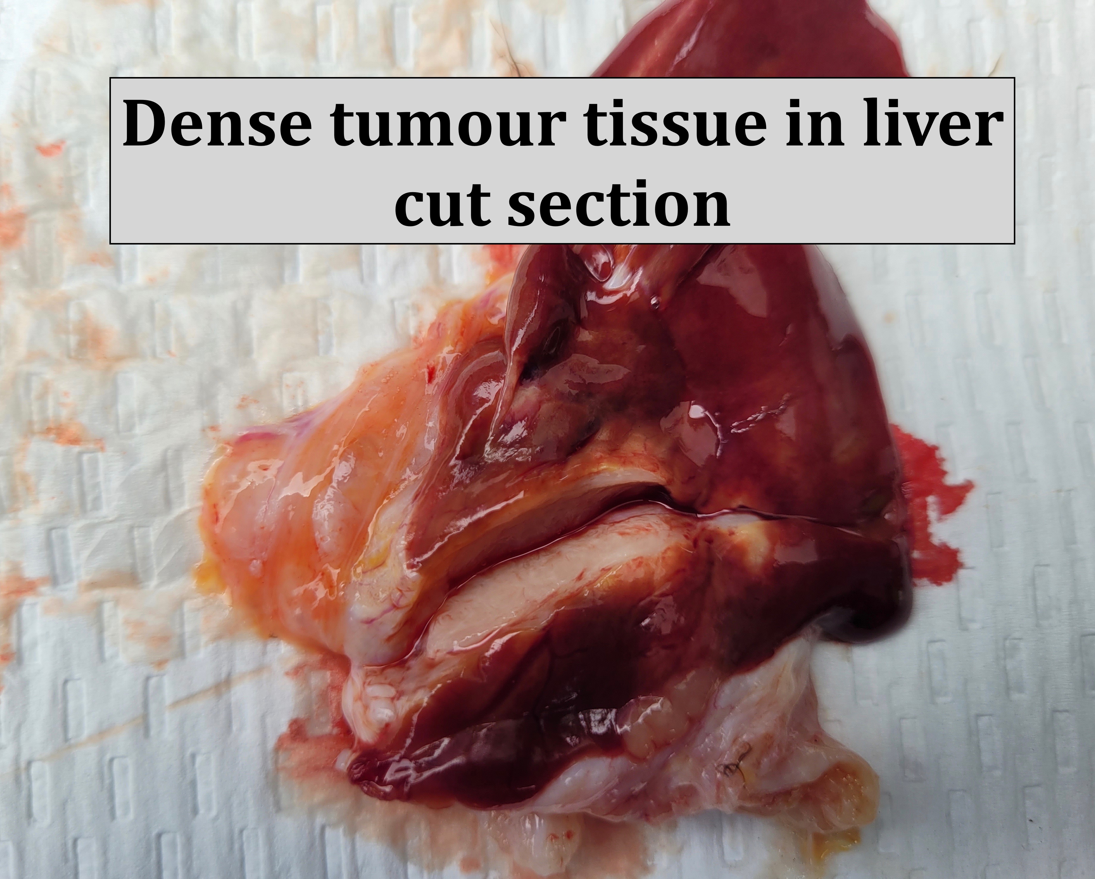
The liver had adhesions making it difficult to lift out. Dr Bowes spent considerable time cutting them to free up the organ.
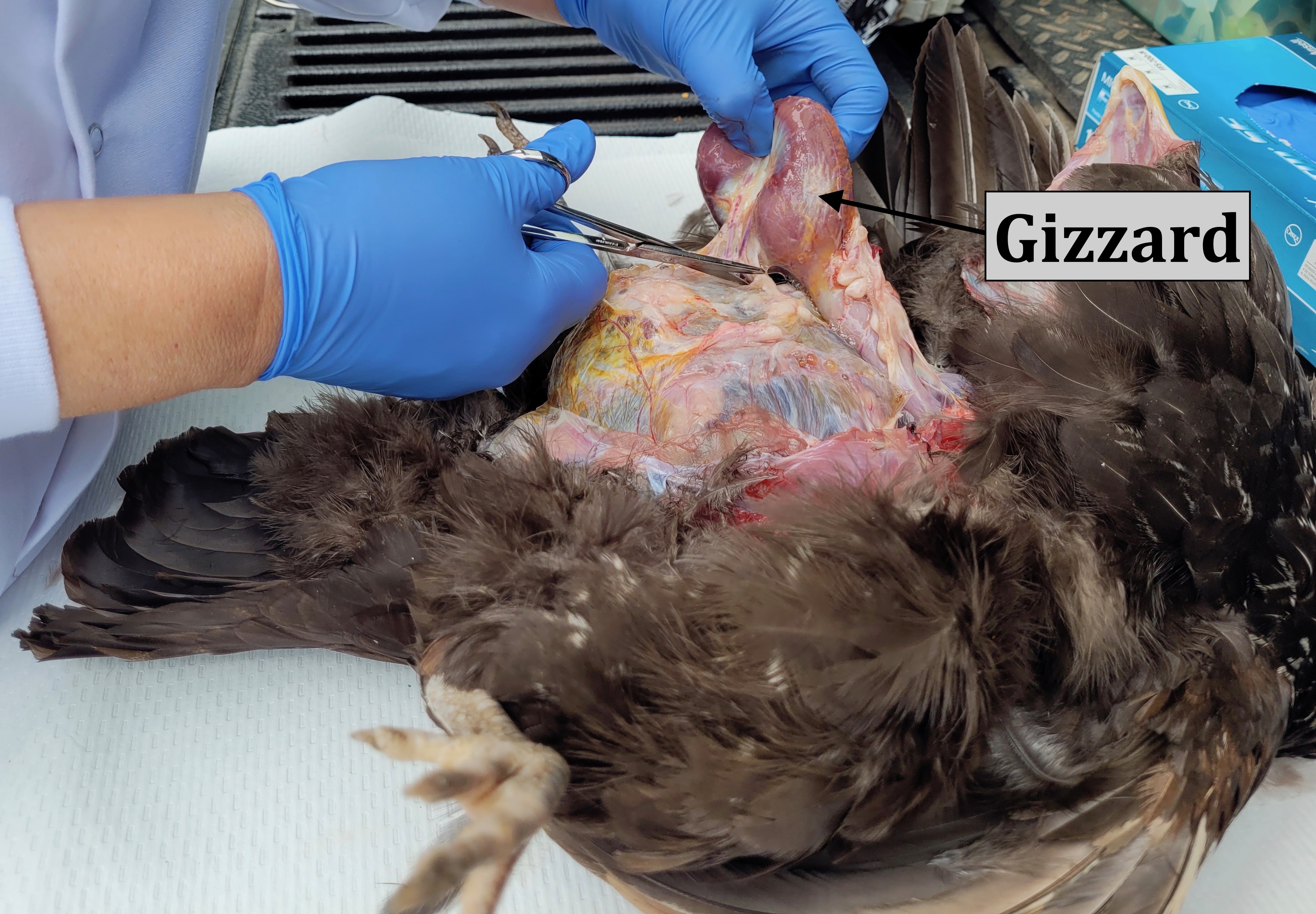
The gizzard was not firm as it should have been but abnormally flaccid, the result of the impairment of nerves. Dr Bowes not only observes how things appear, but feels them. She had me snap on some latex gloves to touch the gizzard.
The crop was full as was the gall bladder indicating that nothing was passing through the digestive system.
There was no fat on the heart, as there should have been; a sign that my hen had used all her fat reserves.
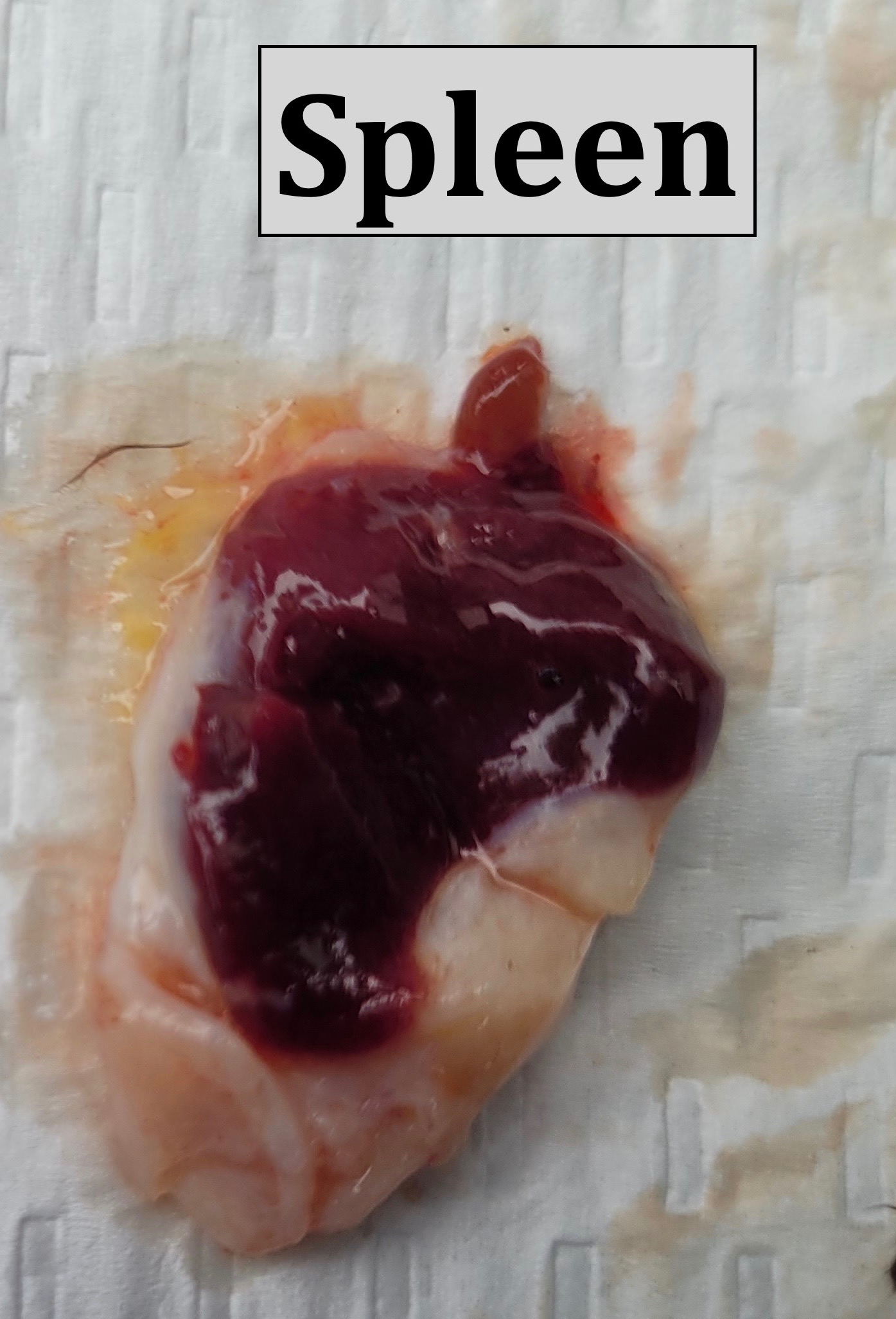
Then Dr Bowes announced that she was ‘tumour girl’, finding one tumour on her liver and others on her spleen, lungs and intestines. In my other cases, Dr Bowes was able to lift out the intestines as one long tubular structure. Molly’s digestive tract was filled with adhesions making that impossible to do. They were also attached to her air sacs, which would have affected her breathing.
Dr Bowes cut her intestines open, which revealed more tumours and undigested feed.

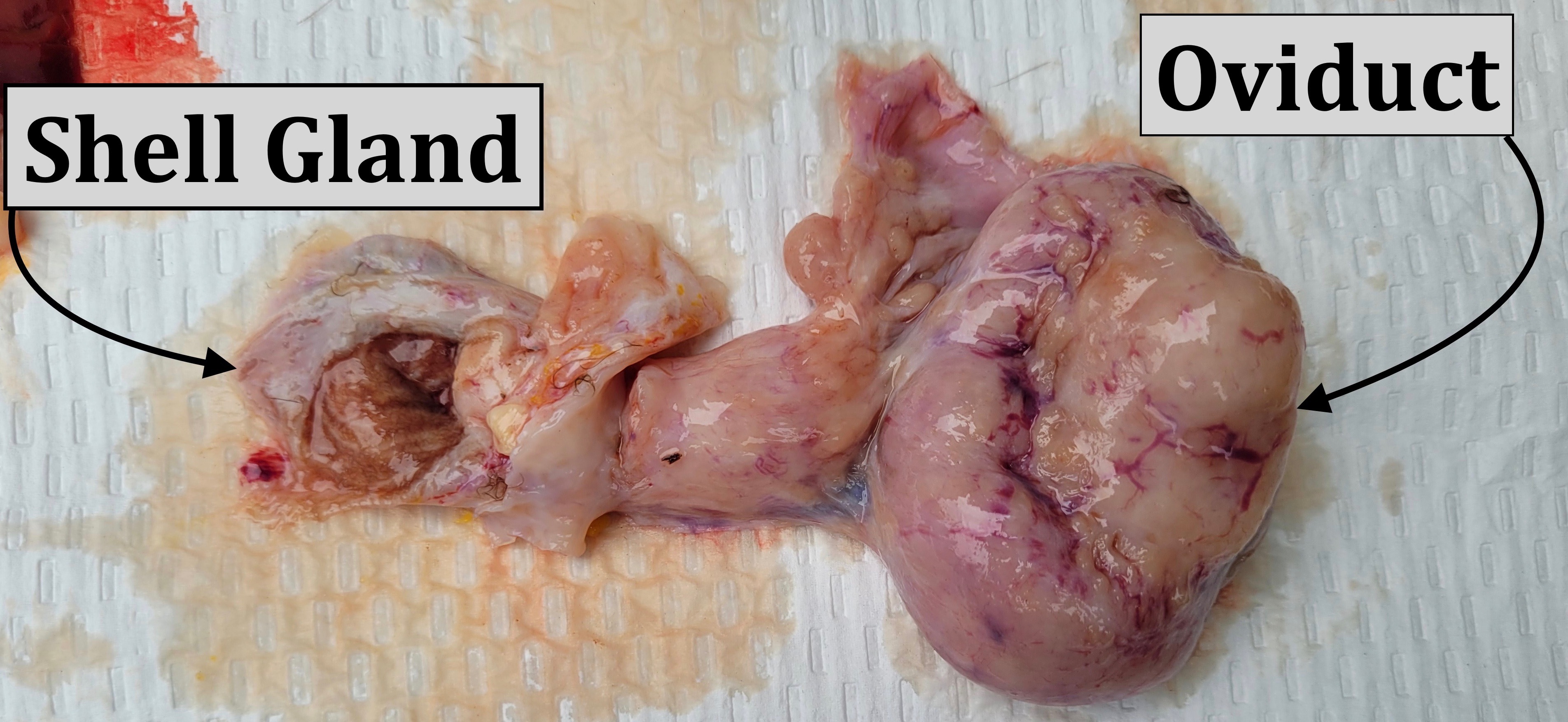


Diagnosis: Ovarian Adenocarcinoma With Abdominal Metastasis
I asked if there was anything I could have done to prevent or treat it, but unfortunately the answer was no. Sadly, domestic hens have been bred to lay an inordinate amount of eggs, which increases their risk of reproductive tract issues including cancer. In this case, her illness was a symptom of a hormonally active organ that inspired an intense fibrous reaction.
FYI: I always encourage folks to keep heritage breeds that have not been bred to lay young, often and large eggs. Production breeds are great layers, but the cost is a shortened lifespan. I also recommend that chicken keepers allow their hens to have long, well-deserved rest periods as egg laying takes a lot out of them. One of the ways to accomplish this is to not use artificial lighting over the winter.
Glossary
Adenocarcinoma: Cancer that forms in the glandular tissue, which lines certain internal organs and makes and releases substances in the body (i.e. mucous, digestive juices, and other fluids).
Adhesion: A band of scar tissue that joins two internal body surfaces that are not usually connected. Organs or tissues within the body stick (adhere) to other internal surfaces. Adhesions develop as the body attempts to repair itself.
Metastasis: The spread of cancer cells from the place where they first formed to another part of the body. In metastasis, cancer cells break away from the original (primary) tumor, travel through the blood or lymph system, and form a new tumor in other organs or tissues of the body.
Tumour: a swelling of a part of the body, generally without inflammation, caused by an abnormal growth of tissue, whether benign or malignant.
Once again, my appreciation goes out to Dr Bowes for generously sharing her time and expertise to further my knowledge of small flock health issues. I try to pass on everything I learn hoping to improve the overall health of small flocks everywhere.

Thank you Claire. And Dr. Bowes. These blogs are so informative and helpful to so many. Never did I realize when I started out with chickens as pets, that they could live such short lives due to various diseases/illnesses.
LikeLiked by 1 person
This was very interesting. Thank you for sharing. It is concerning that this animal had so many tumors. I have learnt a lot through your articles. Keep it up!
LikeLiked by 1 person
Excellent
LikeLiked by 1 person
Excellent. Thank you.
LikeLiked by 1 person
Normally I skim these posts about the diseases as I find them shocking and so sad. I also console myself by reminding me that someone (you) really cares and is making a difference for a lot of other chickens by explaining and sharing the information. Ugly but very informative. Thank you. ml
LikeLiked by 1 person
Thanks for the feedback. I know that a lot of the health cases aren’t easy to read/see, but it’s a great opportunity for chicken keepers to be aware of many of the issues that can affect their flock.
LikeLike
Thanks for sharing this!
I wish all chicken owners would follow you.
LikeLiked by 1 person
That’s my goal 🙂 Sharing my blog is most appreciated. Thanks for the feedback.
LikeLike
Informative piece, thanks for taking the time to post. I’m glad the hen isn’t hurting anymore…poor thing.
LikeLiked by 1 person
I am sorry for your loss. Thank You for posting this very informative article.
LikeLiked by 1 person