Avian Trichmonosis, often called canker (and not to be confused with ear canker) is an infection caused by one-celled protozoa, mostly affecting young birds. It primarily affects the chicken’s upper gastrointestinal system and causes them to develop lesions, or cankers, inside the mouth and the esophagus. As the disease progresses, the lesions get bigger and often interfere with the chicken’s ability to eat and drink. In severe cases, the lesions might block the esophagus causing suffocation.
Its severity depends on a bird’s vulnerability and on the virulence of the parasite’s strain. Chickens that survive may continue to harbour the organism in the oral cavity, crop, or upper digestive tract without showing clinical signs. These birds can shed the parasite intermittently, which is transmitted through saliva, shared waterers, and crop secretions
Adult birds that recover, although asymptomatic, may become carriers and are an important source of infection within mixed-age or stressed flocks.
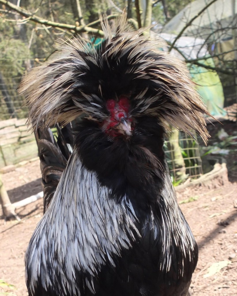
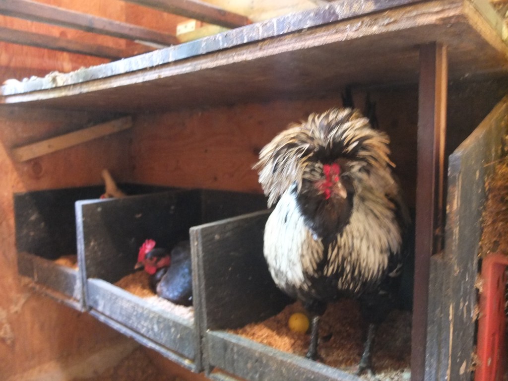
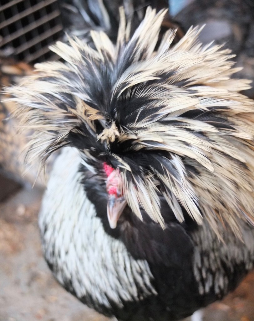
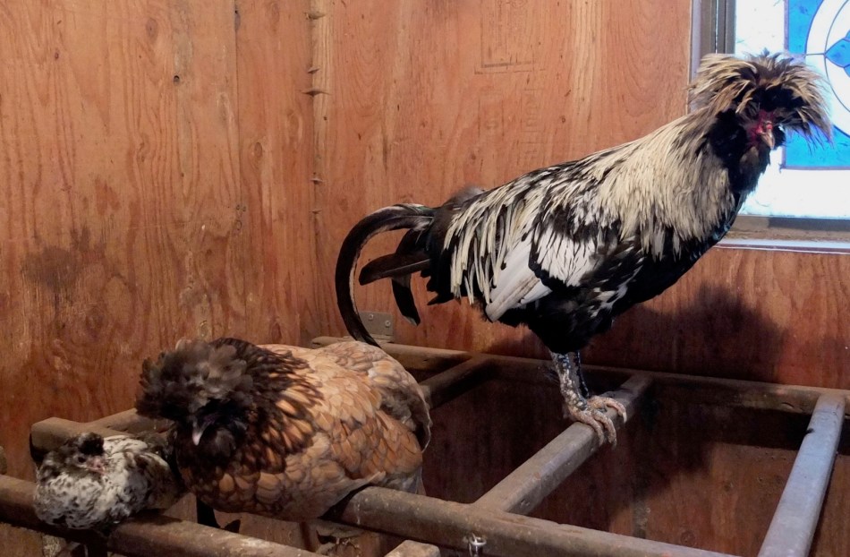
Simon, Appenzeller Spitzhauben x Polish Rooster, 4 years old
I hatched my rooster in June 2017. In spite of other health issues that have affected my flock he’s always been healthy. He’s never been aggressive, but like many crested birds Simon’s a bit flighty and resists being touched so I’ve rarely picked him up.
Recently I noticed saliva coming out of his beak while he was on the roost bar. The next day he seemed okay, but a few days later I saw it happen again. He didn’t have other symptoms and I had seen him eating and drinking, but I thought I should do a health check on him.
Day 1: Simon was in the yard, beak open with some saliva coming out. That must have been happening for a bit because the feathers on his neck were matted. He was shaking his head, scratching his face and making a high pitched staccato squeak once in a while. I found a small hard lump in his neck, the size of a walnut, at the top of his throat (not his crop) and was unsure if it was an obstruction or an organic mass.
His crop was empty, meaning he hadn’t been eating or drinking. I attempted to look in his mouth with a flashlight and didn’t see anything like canker (trichomonosis).
I set him up in a sick bay in the living room with scrambled egg/cat food and water. I don’t think he touched either. He was preoccupied by the congestion in his throat and the mucous coming out of his beak – long gooey strands of it. Simon repeatedly swiped his beak on the shavings to remove it. No coughing or sneezing, but a bit of a wheeze as he was having difficulty breathing.
Day 2: Copious amounts of saliva coming out of his beak. His face and neck were wet with really pungent smelling mucous. The lump was unchanged. I had been attempting to massage the mass thinking it was an obstruction. I could manipulate it somewhat under the skin, but it didn’t change shape or move.
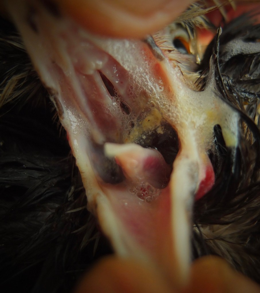
Day 3: He was drinking, but hadn’t eaten since I brought him inside. In 48 hours he only produced one small green, watery poop. Simon continued to drool. I looked in his mouth again, and this time I was able to see a small yellow plaque at the base of his mouth but out of my reach. I did a little research and found that canker can appear in either the throat or crop and not in the mouth.

“Older youngsters or mature birds with a reasonably strong natural immunity will often try to localize a canker infection, leading to nodule formation. If in the throat, these nodules can usually be seen or if in the crop wall can usually be felt as firm mobile lumps ranging in size from 0.5 cm to 4 cm in diameter. Affected birds are treated daily with Spartrix or Flagyl tablets. Once localized (usually 1 – 4 days), throat lesions can usually be teased free with a cotton bud or crop lesions pinched free into the crop. Occasionally, surgical removal is necessary. Premature attempts at removal usually result in excessive bleeding.” – The Poultry Site
As you can imagine this was bad news. Even with medication, treatment would be required to remove the mass in his throat.
Day 4: I called my veterinarian and as I feared this procedure wasn’t in his skill set. I could search for an Avian Veterinarian elsewhere, but felt that multiple visits, medications, and an invasive procedure weren’t practical – especially if it didn’t ensure his survival. I made the sad decision to have him euthanized.
I asked my friend Thomas to help and directed him to do it in such a way as not to damage the mass. He complied and we set about cutting open Simon’s neck. (NB: I wear gloves and encourage you do as well, Thomas opts not to).
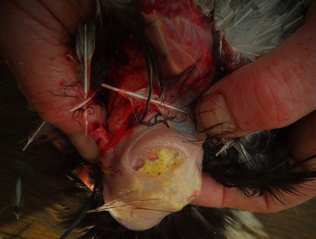
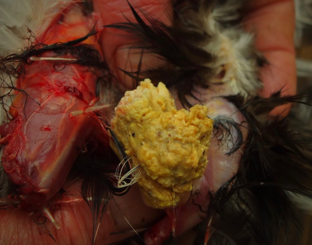

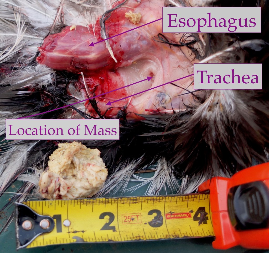
On one side was a large lump visible under the skin. When we opened the area up there was a round spongy yellow ball located at the top of his neck. When extricated it left a deflated pouch of skin. Surprisingly, it didn’t smell.
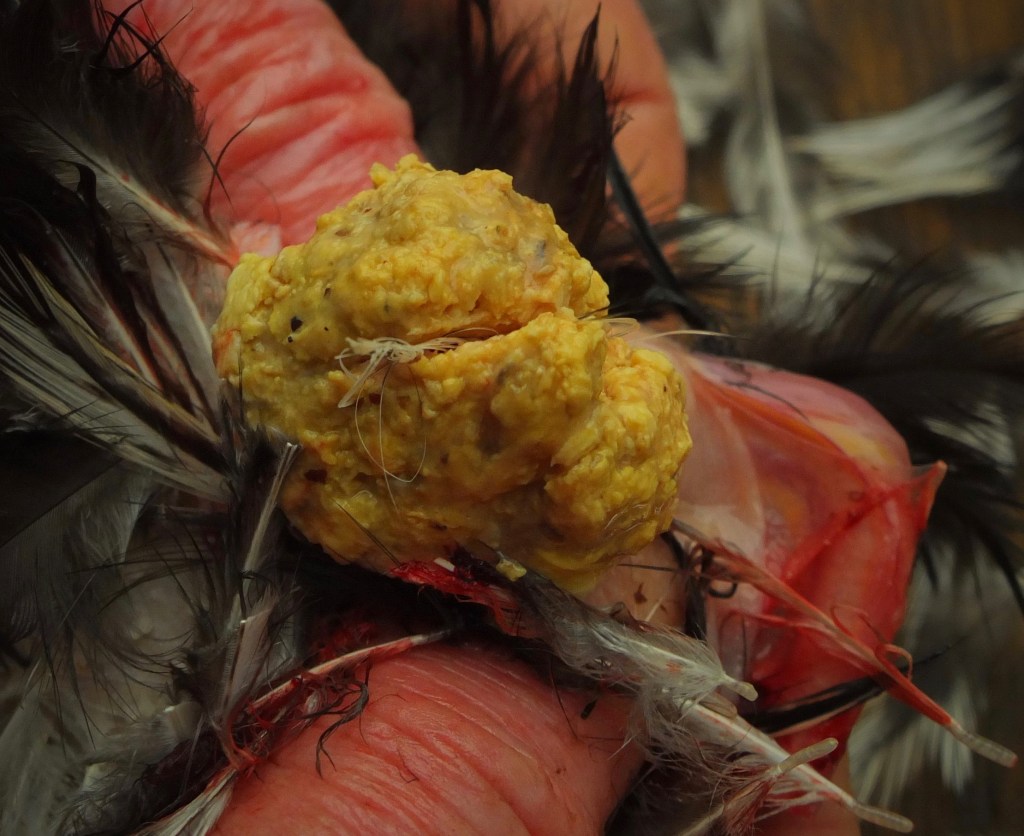
The size of the mass was impressive and scary, in that, it was located at the top of his trachea and esophagus cutting off both his airway and ability to eat and drink. His fate would have been to suffocate or to starve to death. This was a sad decision, but a necessary one as I don’t think there was a way to save him.

We were short on time, so I opted not to necropsy the rest of his body, satisfied that we found the cause of his illness.
I’ve never experienced Trichomonosis in my flock and had some questions that I hadn’t found the answers for online. I sent a series of questions to Avian Veterinarian Dr Jeanne Smith.
At first she was reticent to agree my diagnosis: “I’ve been seeing backyard poultry for 33 years and have only seen a handful of cases in that time – it’s really rare in chickens. Did you look for the organisms under a microscope? If you saw trichomonads the lump would be from ‘canker’. There are mimics of canker: tumors if he was older than 3 years of age or Candida yeast infections. If the lump was found inside the esophagus or crop it could be due to Candida yeast which can cause lumps like that and plaques in the mouth. If the lump was under the skin on the neck or throat outside of the GI tract it could be caseous pus from an abscess or tumour.”
“The definitive test for trichomoniasis is looking at a fresh direct smear of the lesions or the esophagus or crop mucosal lining and seeing the organism swimming around. It can also be definitively diagnosed by histopathology of the tissues fixed in formalin. When I do a necropsy and see lesions like you’ve described I do a direct smear.”
I then sent her the photos that are included in this post to see if she could come up with an alternate diagnosis. “It does look like canker, especially if he also had plaques elsewhere. The lump was in his neck and the mucosa was normal once you removed the lump. The plaques are hard to see, but I think the two together look most like Trichomonas. I can rule out a tumour or abscess. Yeast usually would also cause a “Turkish towel” appearance to the esophagus/crop wall.”
How did he get infected? “It’s most often spread by pigeons, doves or infected chickens that have been introduced to the flock”. I live on a small island off the west coast of Canada where we have no pigeons, very few native doves and I haven’t added new members to the flock since October 2019. I’m also vigilant about biosecurity. It remains a mystery how Simon got it.
How long would it have taken for the mass to reach that size?: “4-6 weeks”.
What can I do to protect my flock?: “10% ronidazole powder put in drinking water – 2 tsp per gallon/water for 7-10 days. There is a 30 day egg withdrawal so you can’t eat the eggs during treatment or for 30 days after treatment. Treating with ronidazole will kill existing trichomonads, but won’t protect them from reinfection.”
Amazon was sold out of ronidazole. I asked my local vet and they weren’t aware of that product and couldn’t find it by that name in their compendium of pharmaceuticals. They did do some research and found that it is not recommended for use in animals designated for human consumption due to the carcinogenic risks. I’m currently looking for effective, but safer options. If you’re interested in more natural options Thyme oil extract has been shown to be effective. Both apple cider vinegar and crushed garlic added to their water once/month are considered a preventative.
Many thanks to Thomas for his unflagging willingness to help out when it comes to the not-so-pleasant side of keeping chickens.
I’m curious as to how ‘rare’ Trichomonosis is in chickens. If you’ve had experience with it please leave a comment or contact me.
Credits: Dr Jeanne Smith DVM; Poultry Site. Featured Photo: Simon, Aurora and their chick in the coop, 2019.
“Serious science. Not-so-serious chickens.”

Just lost a chicken to canker in North Florida. Very sad and scared for my flock.
LikeLiked by 1 person
I only lost the one and haven’t had to deal with it again. Good luck with your flock.
LikeLike
I have a hen who has it now. She is isolated inside and I am treating with a 5-in-1 pigeon tablet. I’ve checked my flock and no other chickens have any signs or symptoms. I’m leaning towards culling my hen because I’ve read it can come back and she can spread it. We live in NW PA and I purchased her as an adult hen and I suspect with the degree in which she had lesions, she had it when I purchased her. It’s just an awful, nasty disease!
LikeLiked by 1 person
Hi, did the pigeon treatment work? I just got an adult hen who has canker (she is separated) and I am not sure whether to keep her or euthanise as I don’t want it spreading, and I don’t want to keep her separated for her whole life. Thanks!
LikeLiked by 1 person
I opted to euthanize my rooster, but I encourage you to treat your hen. Once she’s cured you won’t have to keep her separated.
LikeLike
The takeaway for me here, after reading several of these posts , is to not ignore a chicken who’s having trouble breathing or won’t eat. Either way, it’s probably serious and something needs to be done. Thanks so much, once again, for allowing us to learn from your loss.
LikeLiked by 1 person
My rooster has runny nose, thick gluey saliva in between his beak. Whenever I picked him up thick saliva came pouring out of his mouth and there was bad smell from his mouth. I took him to a non-bird vet; from his symptoms the vet says he has infectious bronchitis and mycoplasma so 5 antibiotic was the treatment. My rooster improved, all symptoms ceased, his appetite increased. On the sixth day he had his eyes closed. 2nd trip to the vet and 8 more days of same antibiotic given. Second day of the 8 day antibiotic he started breathing with open mouth and thick saliva came out of his mouth again. Third trip to the vet. This time he was diagnosed as having thrush and that there plagues of it in his throat so thrush medication. He has stopped eating drinking, eyes closed mostly. When I give him water he wanted to drink but somehow stopped. I checked his mouth and saw yellow soft melted cheese like under his tongue and a long inside his throat in a small vertical sort of long round shape. I used a Q-tip and removed some of it. He still not eat or drink. I don’t know is it canker or thrush. His crops is slow to empty. Thanks
LikeLiked by 1 person
Sorry to hear about your rooster. I’m not sure how the vet diagnosed bronchitis and mycoplasma. I would suspect that it is canker or a bacterial infection. Are you able to get Metronidazole (Flagyl)? It is the recommended treatment.
LikeLike
Have seen it several times….not rare imho, and brutal, despite treatment even in early stages.
LikeLiked by 1 person
I think by the time it’s diagnosed it’s often too late for any kind of treatment.
LikeLike
I currently have a rooster with canker, not responding to meds so far. He free ranges and we did have ringed doves pass through here and I saw a sickly looking little bird on the fence recently. I have tried ACV ACS thyme oil and rotinadazole. Unfortunately none are working and if it’s likely he will be a carrier I’m afraid I’ll have to euthanize him. I thought it was treatable with meds. Sad because he is so sweet.
LikeLiked by 1 person
What does the inside of his mouth look like? Are you able to remove any of the plaques?
LikeLike
I have/had 2 hens with Canker. One needed to be put down. The other, Keri, has been treated with Metronidazole and acidified copper sulfate. She still has it. Tomorrow I will start her on Ronidazole (Ronsec) tablets. 4 tablets the first day, 4 tablets again the third day. I had to call the company to get that info. She weighs 5 lbs. I’m hoping against hope this will work. If it doesn’t I’ll put her down. I’ve been tube feeding her for two weeks with some debridement of the lesions. Not nice for her. Unfortunately, I’m still not entirely sure it’s not wet pox. She has never had the odor in her mouth but I’m not sure that is always present. I read one study which indicated it’s not. I’ll post back with results after the Ronidazole. Meanwhile, do you have any advice about discerning wet pox from canker? She has not had dry pox but from what I’ve read in studies they don’t always go together. Many thanks for your help and informative site.
LikeLike
That’s an interesting question as canker and wet fowl pox both appear as whitish-yellow plaques inside the mouth. Maybe one way to differentiate them is if it responds to a particular treatment or not. I have seen lots about treatment of dry pox but not wet pox.
LikeLike