
At some point, most of us will experience losses in our flock – some expected, others not. It’s always difficult, but more so when you are left wondering why your bird died and if there are any concerns for the health of your other birds. I’m a big proponent of having a professional necropsy done – I’ve sent five birds to the Animal Health Centre – but it is not always easily accessible or affordable for everyone.
This post is not an instructional manual that would replace having a necropsy performed by a veterinarian, rather a guide for how to open a body, recognize gross abnormalities and when cause of death isn’t apparent, to document your finding through photographs and notes for future research.
When you do a DIY necropsy, you’re just searching for obvious issues and won’t be doing the in-depth lab work that comes with a professional necropsy, which would include looking for evidence of parasites and pathogens using a microscope.
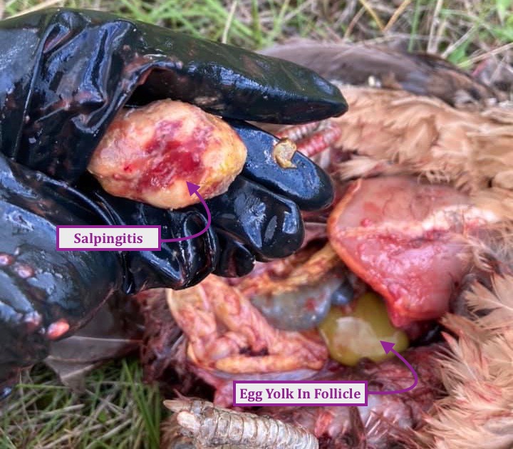
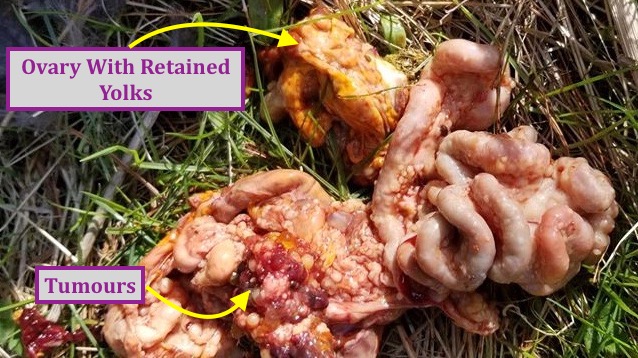
It doesn’t take a vet to see when a bird has died from reproductive issues like Salpingitis, Egg Yolk Peritonitis, Ovarian Cancer or internal laying. You can also find evidence of thickening of sciatic nerves from Marek’s Disease or excess fat accumulations indicative of Fatty Liver Syndrome.
I’ve had two birds that required euthanizing recently. I asked my friend Thomas to do it and then open the birds so we could take a look. He also helped when my friend Tracy’s bird died suddenly. She was going to bury her hen, so I asked if Thomas and I could do a DIY necropsy.
He’s experienced in butchering his own birds to eat, but neither of us has the experience of a skilled dissectionist. He did a rudimentary job, making a basic incision and removing the organs to be photographed. I was glad that he was able to identify what we were looking at and any issues with their appearance. Fortunately, there was enough evidence of pathology that I was able to get a diagnosis by posting the photos online. I’m sure folks were less than thrilled with our lack of finesse, but it got the job done.
Equipment Required
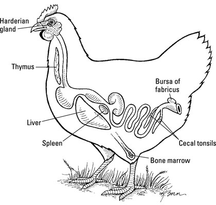
- If you don’t have any knowledge of basic chicken anatomy it would help to download some diagrams or make a quick sketch so you know what you should be looking for
- Level table surface
- Gloves
- Scissors
- Secateurs
- Bucket
- Camera
- Pen & paper for notes
When doing a necropsy you’ve got a couple of options: the basic version that Thomas and I did, which only took a few minutes or a more complete necropsy looking at all the systems of the body that will take longer and require a bit more skill. Remember to take clear photos of the organs in situ and then once they have been removed from the body. Photographs of individual organs and any notes about them are helpful.
Step-By-Step Exam
- Some folks skin a bird completely, others pull out the feathers from vent to keel or just wet the feathers so they don’t blow around.
- Place the bird on its back with its feet towards you.
- Incise the abdominal muscle and cut through the ribs on the sides of the keel bone.
- Make note of any fluids (ascites), odours or excess fat.


- Open the body near the keel end and lift up the rib cage to reveal the first layer of internal organs and the chest cavity.
- You’ll see the liver and heart first.
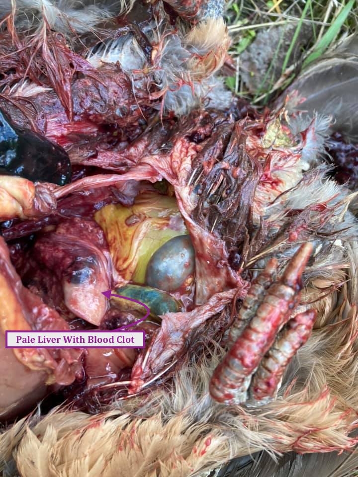
- Examine the liver for changes in size or discoloration, white or yellow spots, abscesses, and/or tumors.
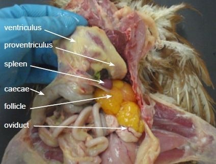
- Remove the liver and spleen. A green discoloration of the liver near the gall bladder is a normal finding. The spleen is the reddish, round organ located at the junction of the proventriculus and gizzard.
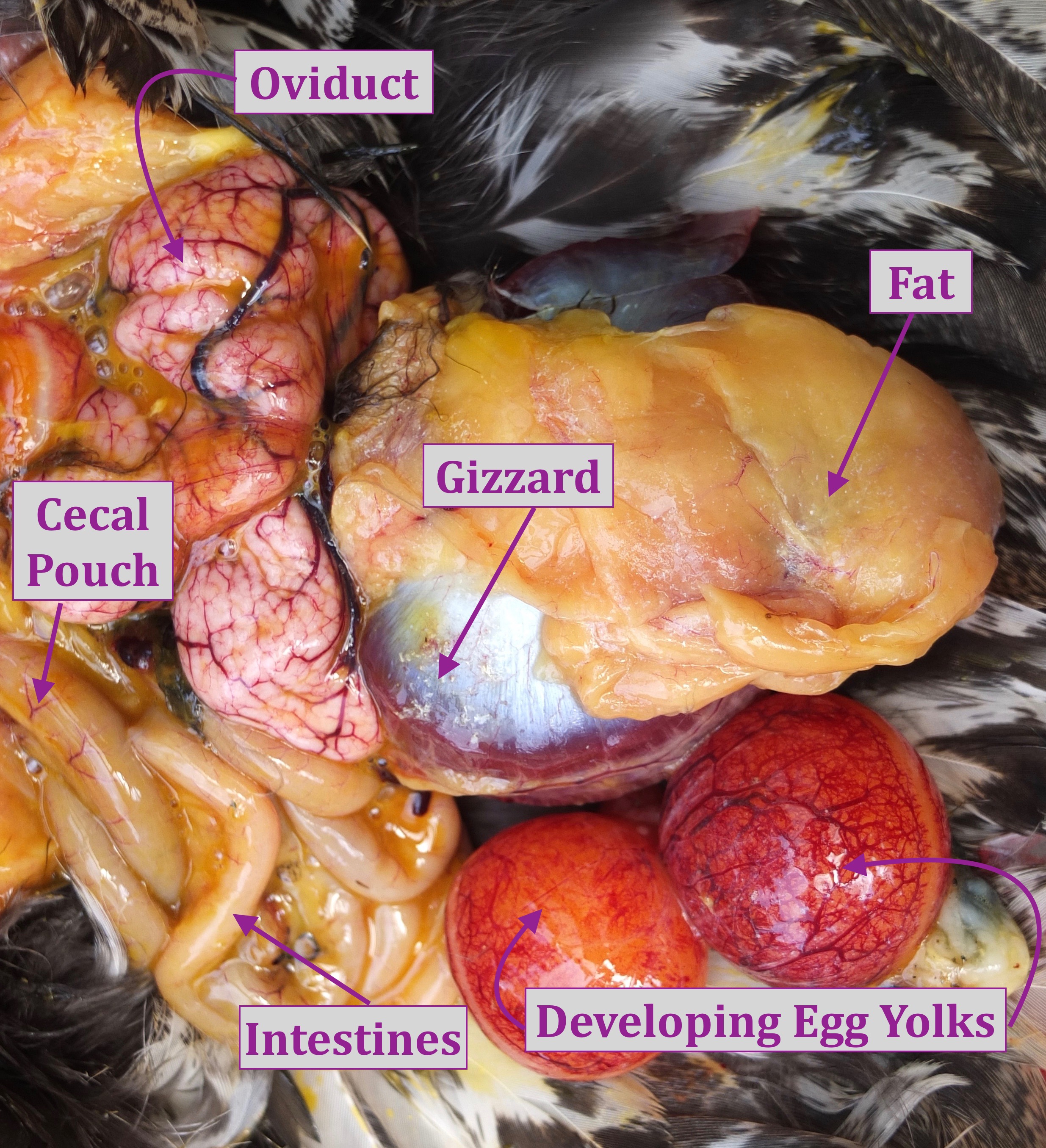
- The next layer reveals the intestines on the left and gizzard mid-right.
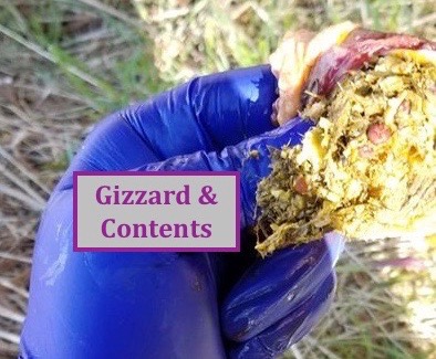
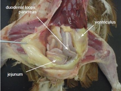
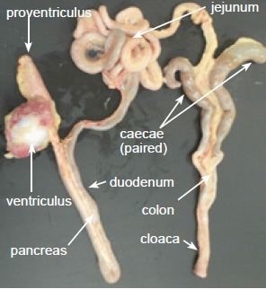
- Remove the proventriculus, gizzard, small intestines, large intestine, ceca, and cut off at the level of the cloaca. The pancreas, the pinkish tan organ within the loop of the small intestine, can also be removed.

- Cut all attachments close to the intestines and set the GI tract aside. At the end of the necropsy, these organs can be opened up and examined for internal parasites.
- The lungs are attached to the ribs at the top behind the spine and the kidneys are tucked along the central spinal column.
- Examine the kidneys, which are elongated, lobed organs that are embedded in the backbone of the bird, and the left ovary/oviduct or paired testes, which are positioned on top of the kidneys.
- The outer surface of the heart should be examined for a cloudy, thickened appearance, suggesting pericarditis. Note if excess fluid is located between the heart and the pericardium (membrane covering the heart).
If you are looking to do a more advanced investigation:
- Expose the sciatic nerve, located on the interior upper thigh (under the muscle) on both legs. The nerves should be the same size with no swellings. Enlargement of this nerve can be an indication of Marek’s Disease.
- To find the bursa of Fabricius, cut through the cloaca and look for a grape-like structure towards the rear of the bird. The older the bird the smaller the bursa, which diminishes in size as the bird reaches sexual maturity.
- Cut the bursa in half. It should have wrinkles running parallel to each other on the surface and be cream colored in appearance. Note any discoloration or swelling.

- Return to the GI tract and starting with the proventriculus, cut lengthwise. The inside wall is bumpy which is normal as these are digestive glands.
- Cut through the gizzard, intestines and ceca. Note the appearance of the inside walls (mucosa) and the presence of parasites (worms), blood, and/or a thickening or discoloration.
- Examine the air sacs for increased thickness and cloudiness. The normal air sac surfaces look like soap bubbles or clear cellophane wrap.
- Cut through the corner of the beak, through the throat and down towards the heart. Examine the interior surface of the esophagus and crop. Look for the presence of food and/or worms in the crop. If the inside surface appears to resemble a towel, it may be an indication of a fungal infection called crop mycosis.
- Cut through the larynx, trachea, and syrinx. The inside surface should be free of excess mucus.
Dispose of the carcass properly and disinfect surfaces and tools.
Thomas and I have conducted several DIY necropsies and each time I learn a little bit more. I also study chicken anatomy and necropsy sites in order to figure out what is normal or not. So far, the cause of death for hens that we have worked on have all been quite obvious: Ovarian Cancer, Egg Yolk Peritonitis, Fatty Liver Disease and Internal Laying. I won’t have the full story that a professional necropsy provides but an at-home diagnosis gives you closure and peace of mind.
It’s important for most of us to know that:
- our bird wasn’t unnecessarily euthanized
- the health issue isn’t likely to be transmitted to our other birds
- other flock members with the same symptoms can be euthanized or treated sooner to prevent unnecessary suffering
If you’ve got some necropsy photos and some case notes you’d like to share with me feel free to drop me a line.
Credits: Government of Western Australia (Agriculture & Food); The Poultry Site; Kendel Alstad and Crystal York.

Curious, what is the organ shown in the photo that shows in the photo immediately after this phrase “Make note of any fluids (acites), odours or excess fat.” (Unsure how else to identify which photo I’m looking at, lol).
I’ve just seen the same thing on a quick necropsy (a thin fluid filled, vascular sac) that didnt appear to be part of the GI tract. I’ve done necropsies quite a few times and have never come across something similar (though, to be fair, it’s mostly roosters I’ve dealt with).
LikeLiked by 1 person
That’s a good question. The hen was my bird, but I don’t have a confirmed cause of death. It’s difficult to tell from the photo exactly what we’re looking at. It would help if the photo showed where it was located in relation to the other internal organs. I asked a vet tech friend and she suggested it might be a fluid filled cyst. If anyone else has input I’d be open to hearing it.
LikeLike
Prompted by your mention of “chicken anatomy and necropsy sites” I went to YouTube. It hadn’t occurred to me for some reason. I go there for everything else. Looks like Cornell has what I want. Thanks 🙂
LikeLiked by 1 person
PS All I need now is a real sick chicken! I just like being prepared.
LikeLiked by 1 person
If you ever do a DIY necropsy send me some good photos and notes and I’ll show them to Dr Bowes, my avian vet/pathologist.
LikeLike
Very interesting stuff here. I thought the guide was very helpful and I appreciated the explanation at the end. Many people may find this hard to do, but some of us want to know.
LikeLiked by 1 person
Even if folks can make the incisions and take photos of the internal organs they can then post them online for help (or send them my way for help with a diagnosis).
LikeLike
That’s very kind of you. And posting pics online can be extremely helpful to other chicken keepers who are also searching for answers. That’s something we should keep in mind. Our loss can be turned into something that’s beneficial down the road
LikeLiked by 1 person
I want to thank you. Here in 2025, I culled my first chicken and I was very heartbroken but I needed answers, she had stopped eating, she stopped clucking, and I didn’t want her to suffer anymore. This was very helpful and I may be reaching out to get some insight into what I saw/have photos of. Thank you again this helped immensely.
LikeLike
Dealing with end-of-life issues is never easy, but having some answers about what was going on with her health might give you some peace of mind. Thanks for the feedback.
LikeLike