
Several months ago Dr Vicki Bowes, Avian Vet/Pathologist offered to meet up with me on a regular basis to help me figure out the cases in my ‘What Is This?” file. We’ve been going through case notes, necropsy reports and photos to reach what Dr Bowes terms her ‘best guess’. We recently spent a marathon 4½ hours sifting through 27 mystery cases wanting to be solved. Most of them were old hat for her, although there were a few that presented some challenges. To see the ones that tested her more than 30 years experience as an Avian Pathologist click here.
I’ve grouped them by type. This file is called ‘Lumps & Bumps’, a medley of all kinds of wounds, cancers, lesions and more, just waiting for Dr Bowes to give me her diagnosis. Again, she did not disappoint and I present them here for your learning.
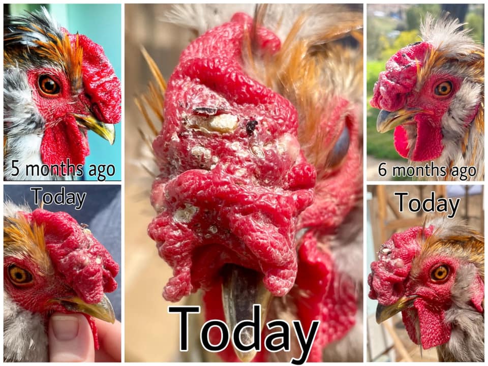
Bumps On Neck

Dr Bowes was pretty confident she could rule out Favus (ringworm), but had she dealt with the patient in person she would take a skin scraping for diagnosis. In the absence of that information, she suggested this is a fungal infection and suggested treatment with the over-the-counter product Tinactin that is recommended for Athlete’s Foot.
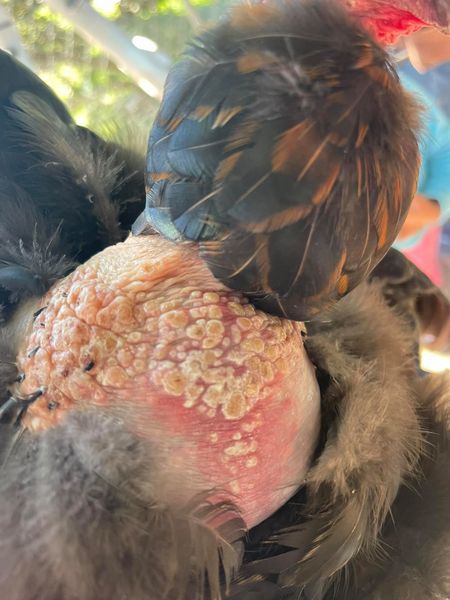
Pedunculated Caruncle

This case was presented as a growth from the hen’s ear, which it is not. Nor is it Fowl Pox or an ear infection. In fact, it is adjacent to, but not originating from, the ear. It’s an abnormal caruncle (i.e. a fleshy growth like a wattle). Dr Bowes’ concern was if the growth got larger it might get abraded leading to infection or it could occlude her ear/auditory canal. She suggested this was not an opportunity for DIY surgery, but recommended that the bird should be taken to a vet if it became a problem.
Lump On Leg #1 (Tiffany Maxwell)

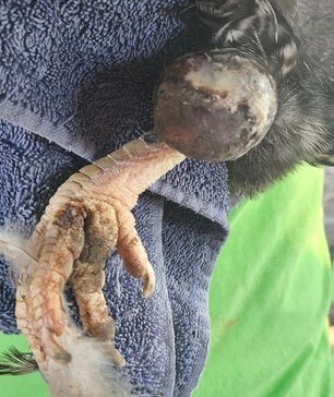
Dr Bowes suggested that this lump was too discrete (i.e. distinct edges) to be a foreign body reaction, but was most likely to be a localized, neoplastic tumour (abnormal tissue mass). Short answer: this is skin cancer, which would require surgery to remove, and even then, there would be no guarantee that the removal of the lump wouldn’t damage her leg.
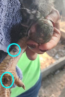
Lump On Leg #2
This chicken keeper had a hen that presented with a lump on her leg. She was treated with antibiotic ointment and appeared to be getting better although the leg was still discolored and swollen. She died several weeks later. Subsequently a second hen presented with a lump on her leg.


Dr Bowes: The lump is ulcerated and open with a localized infection. The black area needs to be addressed and treated as a wound: cleaned, debrided, given antibiotics and covered. This is a case that can be resolved through dedicated, consistent supportive care.
Bitchin’ Chickens: My two cents is to have a vet prescribe Clavamox in this case.
Lumps On Body
“I just got a group of three hens from a friend to add to my flock. This one was limping for a week before they rehomed her. I found crusty patches all over her body, which come off easily like a scab.” – Nya Gawlikoski


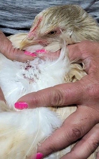
Again, we would have liked better photos, specifically before and after cleaning the affected areas. When Dr Bowes saw the first photo she suspected it was an impacted feather cyst, but then became concerned when there were multiple sites involved. Two things of note: the area was moist in appearance and the white proliferative tissue was abnormal, pointing to squamous cell carcinoma, a form of skin cancer. The lump will continue to get larger and is vulnerable to infection. The only way to address the situation is through surgical removal (or euthanasia).
Skin Issue


Sometimes with photos we’re not always sure what we’re looking at. It appears the area in the first photo is also captured on the left hand side of the second photo. Dr Bowes noted the raised area of squamous cells (outer skin) and infiltrative tissue (i.e. the diffusion or accumulation in tissue or cells of foreign substances or in amounts in excess of the normal). In this case a biopsy would be warranted to determine if the skin is infected and if so, how deep or is there a tumour? Is that feather loss normal molting or a is the bumpy skin a reaction to feather loss?
Spots On Comb
“Smudge is a 1.5 year old Silkie cross with a walnut comb. He has occasionally had small areas of slightly powdery white on his comb, which I would wipe with Vetericyn. I figured it was just due to the folds in his comb and it never looked inflamed. Today I noticed what looks to be some kind of infection.” – Amber Mersino
The white powdery material is suggestive of a yeast or fungal infection called Favus, although it could be Candida albicans. The remedy is a combination ointment that treats both fungal and bacterial infections, either Tresaderm or Panalog.
Lumps On Chick Hocks
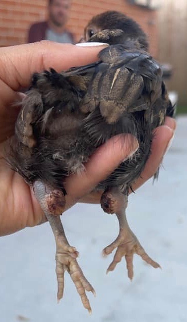
Even though this chick is still very young, Dr Bowes thought the lumps were caused by chronic irritation similar to bedsores. To make a proper diagnosis she would need to know what caused that irritation; how long the chick has had those lumps; is its conformation normal; and does it walk normally. Her recommendation was to humanely euthanize the chick.
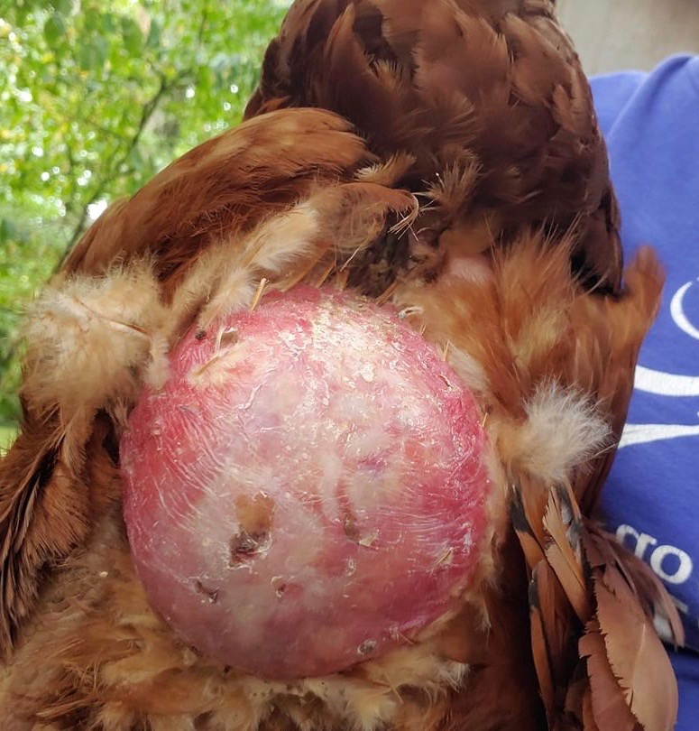
Lump On Crop
Here’s another case without much of a back-story. The tissue is gangrenous and necrotic, not just at the surface of the skin, but into the crop. Because of the depth of infection and the probability of bacteria entering that wound Dr Bowes recommended humane euthanasia.
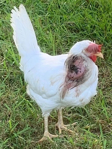
Lump On Abdomen
This is a discrete (defined edges), contained lump. The pale tissue speaks of a neoplasia (abnormal mass of tissue), like cancer. Dr Bowes suggested it could be a chronic breast blister or that a foreign body, like a sliver, created that response. Her prediction is the lump would be draining the hen’s resources, who would be thin despite her weight. A biopsy would be required in order to diagnosis properly, but Dr Bowes recommended humane euthanasia.
So that wraps up 10 short stories from the “Lumps & Bumps” file, not many with happy endings.
Our experience did suggest a few things: 1) monitor your birds on a regular basis and look for help, sooner rather than later; 2) when a case is serious consider humane euthanasia to end your bird’s pain and suffering and 3) regardless of whether you seek online peer assistance or in-person veterinary care it helps to have good notes with information about timelines, symptoms and treatments. Take as many clear photos as you can.
If you undertake a DIY necropsy make notes and takes lots of good photos. Dr Bowes uses both her nose and fingertips to help her make a diagnosis: note any unusual odours and feel everything and report on texture, weight and consistency so someone with more know-how can put the pieces together.
Again, many thanks to Dr Vicki Bowes for generously offering her time and expertise to help us all become better small flock keepers.
If you’ve got a burning question or some photos relating to a chicken health issue send them my way via the contact button on the home page.
Featured Photo credit: Dr Vicki Bowes

I am very interested in all your Avian Pathology posts…there should be more sources for real info like this!! Astounded by how far people let various conditions go though. Interesting. Thank you both for your efforts. Emma
LikeLiked by 1 person
Thanks for you feedback. Dr Bowes are getting together again next week, so there should be a whole new bunch of pathology cases posted soon.
LikeLike
Rough. Poor birds.
LikeLiked by 1 person