
Lymphoid Leukosis is the most common type of cancer caused by the Avian Leukosis Virus. It can be spread vertically (from the hen to the chick before the egg is laid) or horizontally (between flock members).
Tumours usually affect the liver, spleen, and bursa of Fabricius and, less commonly, the kidney, lung, gonad, heart, mesentery, and bone marrow and are often not detectable until 14 weeks of age. Death rarely occurs before then and is more frequent around the time of sexual maturity.
Leukosis can be mistaken for Marek’s Disease, another virus that causes tumours. While Marek’s primarily affects birds under four months of age, Leukosis is slower to develop so usually occurs in adult chickens.
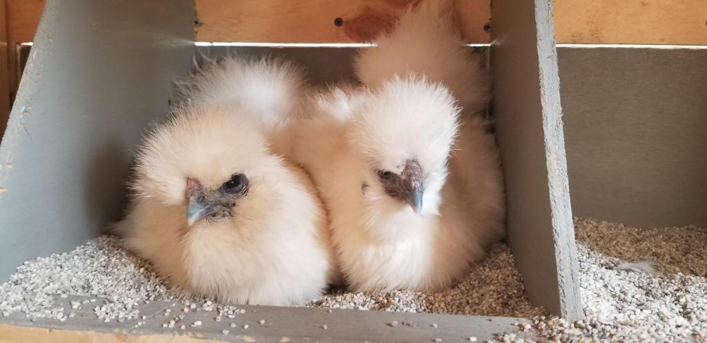
Nugget, 18-month old Silkie hen
Bitchin’ Chickens follower Lara sent me her DIY necropsy photos and notes eight months ago wondering what had killed her hen. I recently showed them to Dr Vicki Bowes, Avian Pathologist for her opinion and used her comments to label the photos.
Lara sent me the following information documenting the timeline of Nugget’s symptoms:
“I am attaching the pictures, with the knowledge that I didn’t take them correctly. I should have laid out the organs so it was clear what they were.
Nugget was sick for about a month before passing away, with symptoms that included:
- Weight loss
- Lethargy
- Stood puffed or hunched position by herself
- Green, gritty and sometimes slimy diarrhea
- Crop was intermittently full
- Toward the end of her life I felt a hard, somewhat malleable mass in her belly. At first I thought it was an egg, but it became clear that it was an organ or tumor.
- Stopped laying eggs
No signs of:
- External parasites or visible worms
- Respiratory illness
- Neurological issues
- Difficulty laying eggs
- Wryneck
Necropsy observations:
- Her intestines were completely blocked with clay-like poop
- The portion right near the cloaca was bloated with diarrhea
- I did not find a lash egg or developing eggs
- The gizzard looked to be filled with the all-purpose sand that I have on the floor of their run. I was also feeding her egg yolk by mouth before death, so maybe that’s why it’s so yellow in there.
Dr Bowes diagnosed Nugget as having Lymphoid Leukosis and noted that there were numerous tumours: soft, white and irregular ones throughout the liver; a large tumour in the spleen; and tumours on the gizzard and cecal pouches.


Lara asked: “Are the small, white hard tumors different from the large, soft ones that are almost part of the organ itself?” Dr Bowes replied: They might be the same type of tumours. If they were necrotic the middle becomes hard. It’s difficult to know for sure without examining them”.
Many thanks to Lara for sharing her story and photos and to Dr Vicki Bowes for so generously sharing her expertise.

0 comments on “Case Study: Lymphoid Leukosis”