
After 13 years of keeping chickens and many hours scrolling through online poultry sites I’ve seen my share of oddities. Since starting this blog I’ve been keeping a file of cases that I’ve read about or Bitchin’ Chickens followers have sent to me. Sometimes I know the diagnosis, but other times I’m stumped and relegate them to the ‘What Is This?’ folder.
I’ve had the good fortune of spending time with Dr Vicki Bowes, Avian Vet/Pathologist lately. She invited me to sit down with her to look at the DIY necropsies and oddities. Some of them were accompanied by clear photos and good notes and I’ve been able to turn them into case studies. Others were snippets with not enough material to write a full article but interesting, so I’ve decided to present them here as short stories as a learning experience for those of you curious about chicken health issues.
Full credit goes to Dr Bowes for her expertise; I’m merely the one who collates all the material. FYI: These diagnoses are made to the best of her ability without the benefit of seeing the birds in person.
Odd Structure?


Bitchin’ Chickens follower, Charna sent me this: “I found this structure directly behind the vent of a 12 week old cockerel. I’ve been processing my cockerels for a few years now and never noticed this before.”
Dr Bowes: That’s the bursa.
Bitchin’ Chickens: For further explanation I’ll add that the bursa of Fabricius is a lymphoid organ unique to birds and a critical part of the immune system where the B-cells mature. When an antigen is encountered, the B.F. programs these cells to attach to the antigen then move to the blood, spleen, cecal tonsils, bone marrow, Harderian gland and thymus. Destruction of the B.F. in a young bird (e.g. by Infectious Bursal Disease or Marek’s Disease) prevents the programming of B-cells for the rest of their life. Affected chickens won’t be able to respond to diseases or vaccinations by producing specific antibodies.
Bone Cysts?
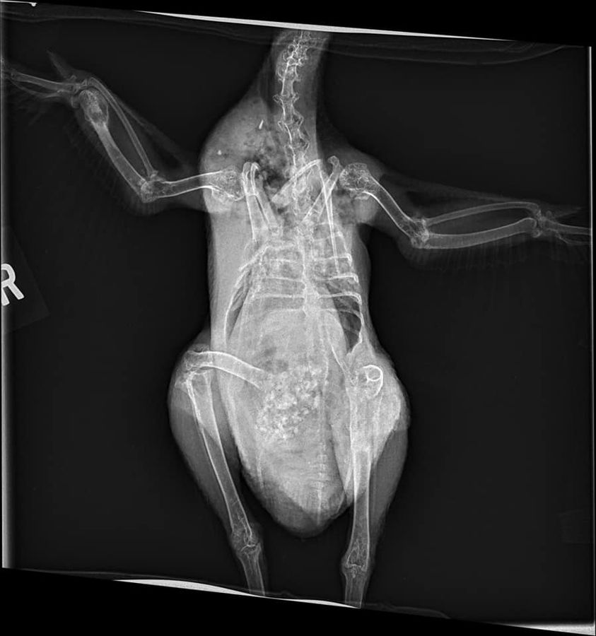
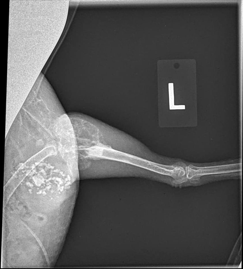
“Our Silkie hen has had a limp that has been getting worse over the last year. The vet took x-rays and sent them to a specialist who diagnosed a bone cyst in her leg and on her wing. She is now on Metacam, but we’re unsure where to go from here. The vet says we could do a biopsy of the cyst, but that would be in the $600-700 dollar range and we can’t afford that right now.”
Dr Bowes: The areas of deterioration in the wing and hip are not bone cysts. Osteosarcoma is one condition that eats bones, but is rare. Avian Tuberculosis is another possibility, but this case is more likely to be linked to bacterial arthritis caused by a Staphylococcus infection. There are multiple sites of deterioration in the wing (distal ulna) and no bone articulation in the hip making it increasingly difficult for the hen to walk. Recommendation: humane euthanasia.
Abnormal Leg?


“This is the only chick of eight with this condition. I have tried Neosporin, Lamisil and Vetericyn Plus. Nothing seems to help. In addition to the dark crusted area the scales on her leg are missing and the skin is very smooth. She is walking with a limp.”
Dr Bowes: The leg has been degloved and the skin sloughed off, probably due to an injury at the hip which has prevented blood flow causing vascular impairment and possible gangrene. Essentially the leg is dead. This isn’t an injury which will heal or improve. Recommendation: humane euthanasia.
Tumours?

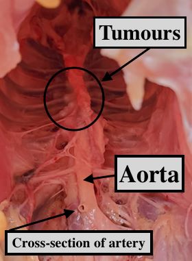
Another Bitchin’ Chickens follower, Chantal Claire sent me this case: “My six-year-old Rhode Island Red hen stopped laying one or two years ago due to age, no health issues. I butchered a number of cockerels and older hens (including this bird) and when I opened her up found tons of these little bubbles or tumors along her spine. Any idea what they are?”
Dr Bowes’ diagnosis: Oviduct carcinoma (you can see the tumours on the oviduct as well as along the spine).
Many thanks to the folks who asked for my help and to Dr Vicki Bowes who, once again, so generously came to the rescue in providing the answers.
If you have an unsolved mystery feel free to send your photos and notes to me and I’ll see if I can track down the solution.
Featured Photo: Dr Vicki Bowes at her microscope.

That “degloved” leg is horrible!! Poor thing
LikeLiked by 1 person