
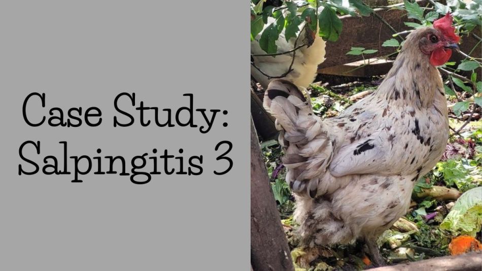
Lucy, Isbar x Black Copper Marans Hen, 22 months old
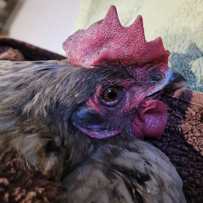
When you have a flock of 30, it’s somewhat easy to miss a bird that is a little ‘off’, especially since they are designed to hide their vulnerabilities and any signs of weakness. Given the changing weather and increased hours of daylight I’ve been spending more time with my birds. Everything looked pretty good, fingers crossed. A couple of days ago, I noticed one hen walking just a tad more slowly – nothing dramatic like the upright stance of a penguin or dragging her abdomen on the ground. I felt her abdomen and thought it was a bit full. I wondered if it was ascites (water belly) and prepared myself to drain a bit to provide her with some relief. I wasn’t convinced that would be anything more than a short-term fix, but I wanted to give her a chance. Notice the tips of her comb are turning blue? That’s cyanosis, an indication that her heart is not pumping oxygen to her extremities.
I set up my kitchen counter, covered with a towel, and my hen swaddled like a tortilla with her head covered. I attempted the procedure three times with no success at draining anything before realizing I misdiagnosed the situation.
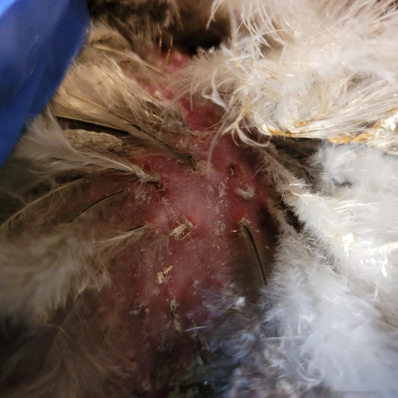
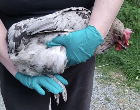
Two days prior I was sure her abdomen was soft and squishy and now it felt hard. Bad news, as that is a sign of a mass (which could be several things including cancer or egg yolk peritonitis). I called up my chicken friend, Christy and asked if she would euthanize her for me. I took a workshop on humane euthanasia, but that was 11 months ago and my skills have been untested. Frankly, I was not confident I wouldn’t screw it up and I knew Christy had loads of experience.
I had her look at Lucy prior to euthanizing her and we agreed that the abdomen felt taut and distended. Nothing to be done about that. It went off without a hitch and I left with my dead hen in tow. In the back of my head, I was sure it was salpingitis.
What Is Salpingitis?
Salpingitis, an inflammation of the oviduct, can be caused by fecal bacteria that migrate up the oviduct or respiratory infections that move down into the oviduct. It’s often associated with Mycoplasma and bacteria like E.coli and Salmonella.
Some of the symptoms are so generalized they could be many things: lethargy, loss of appetite, ruffled feathers, yellow poop or respiratory illness. An affected hen will eventually lay fewer eggs or stop laying altogether. Other typical signs include distended abdomen, laying soft or wrinkled eggs.
All the components of unlaid eggs accumulate and fester in the oviduct, filling it with a smelly, rotting mass. The tell-tale diagnostic clincher is the production of a ‘lash egg’: the passing of an egg-shaped mass of hardened pus, tissues and bits of unlaid eggs. If the hen is unable to pass the mass it will continue to accumulate material and may affect her ability to breathe.
Ordinarily I would put specimens in my freezer and then have Dr Bowes do the necropsy with me. No such luck – my freezer was full and Dr Bowes wasn’t available. I put her in my egg fridge and determined I would do a DIY necropsy on my own the following morning. When Dr Bowes and I get together she does all the skilled work while I ride shotgun: taking photos, making notes and asking questions for clarification. Unfortunately I was on my own and had to multitask.
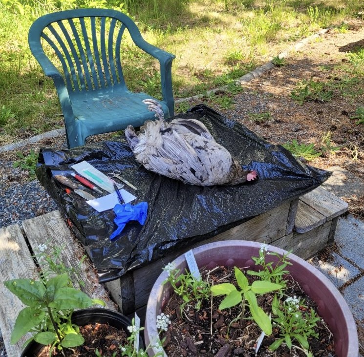
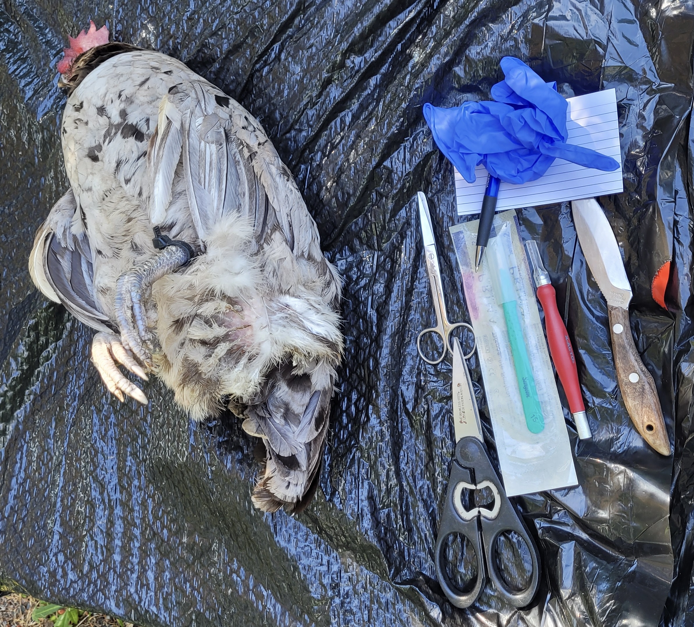
I set up my workstation on the platform to my now-gone hot tub, covered it with a garbage bag and laid out my tools: a knife, scalpels, scissors, camera, pen and paper. I donned my gloves and set about investigating her cause of death. When you watch a seasoned pro like Dr Bowes it all seems so easy. What I discovered was both my scissors and knife weren’t as sharp as they should’ve been. I cursed because I’d already started so wasn’t going to stop to find more effective equipment. The old Boy Scouts adage ‘Alway Be Prepared’ kept repeating in my head.
I stripped off some breast feathers, made an incision down both sides of her abdomen and attempted to pull the keel upwards in order to expose the internal chest cavity. I say ‘attempted’ because it was a beast to get it to release.
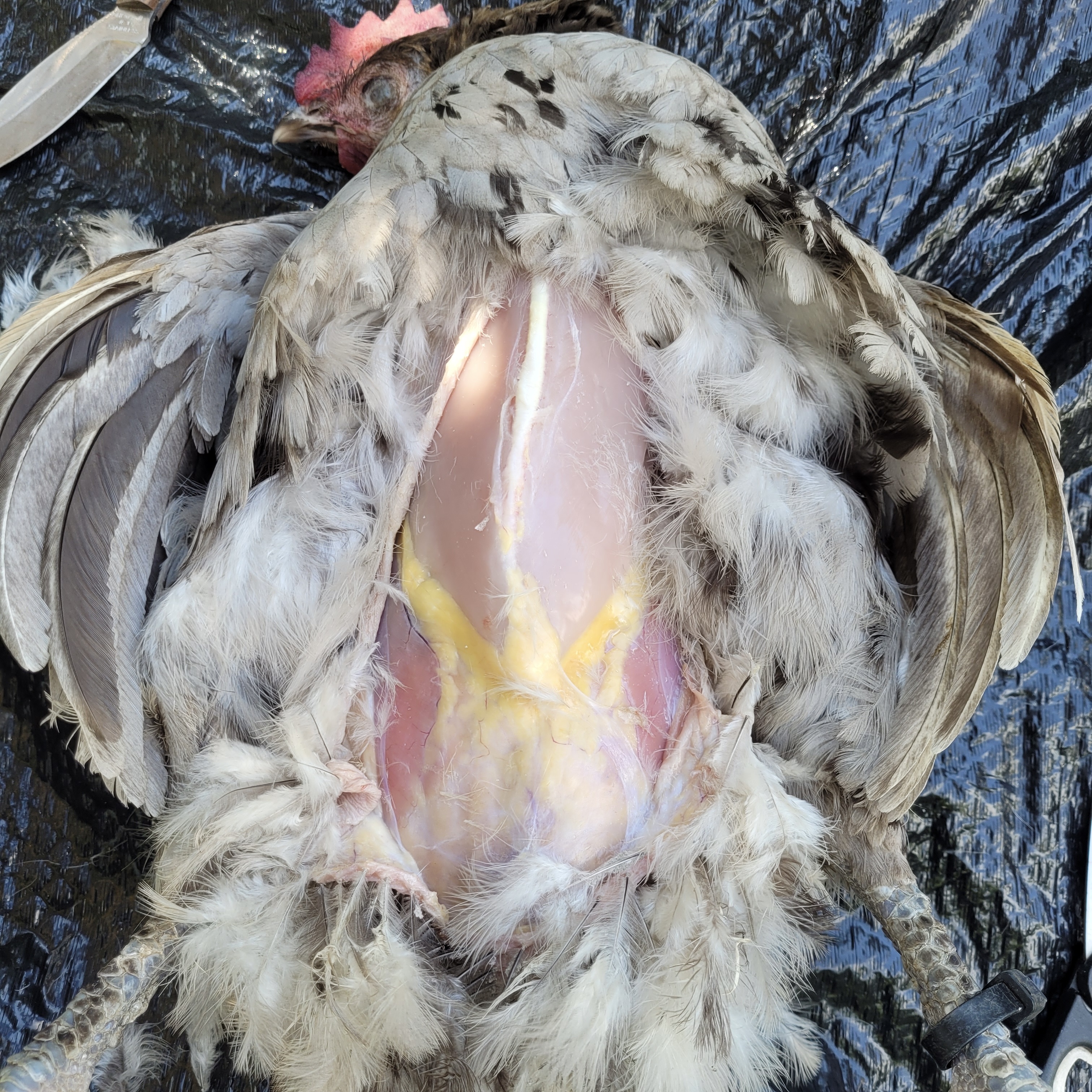
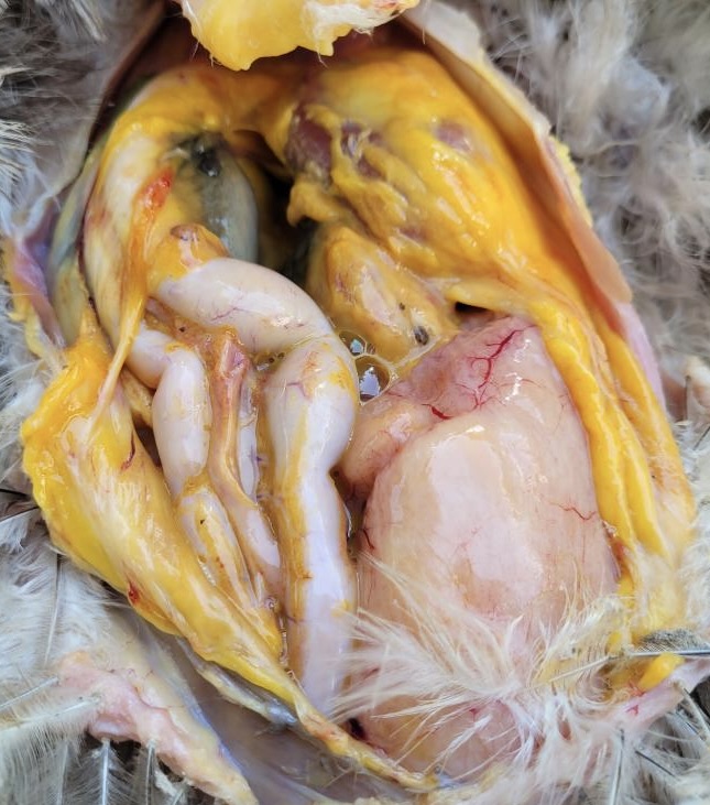
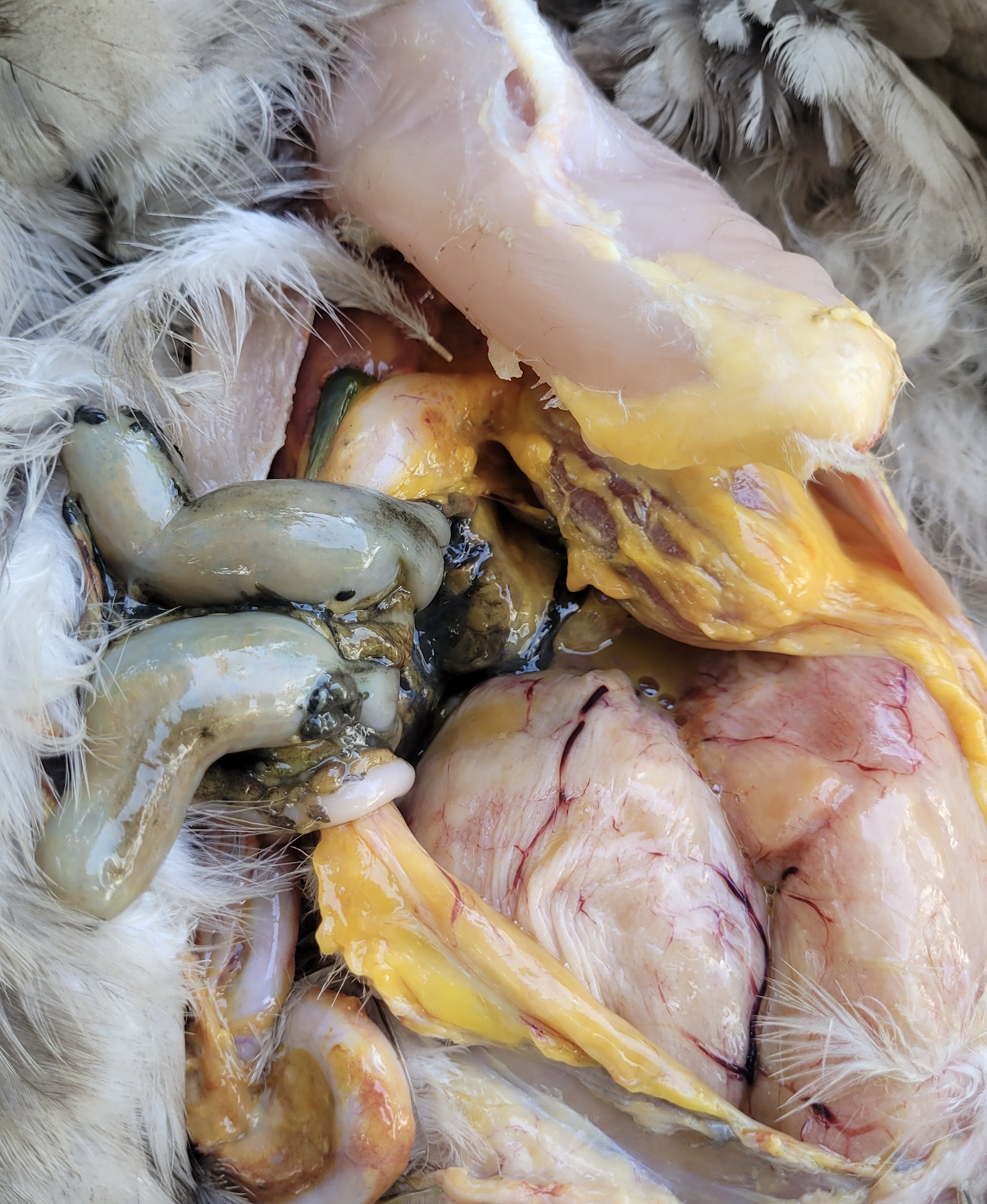
What did I discover? The liver was pale and friable, which pretty much fell apart when I tried to remove it. There was evidence of developing egg yolks. Her gizzard was full. Overall body condition: good.
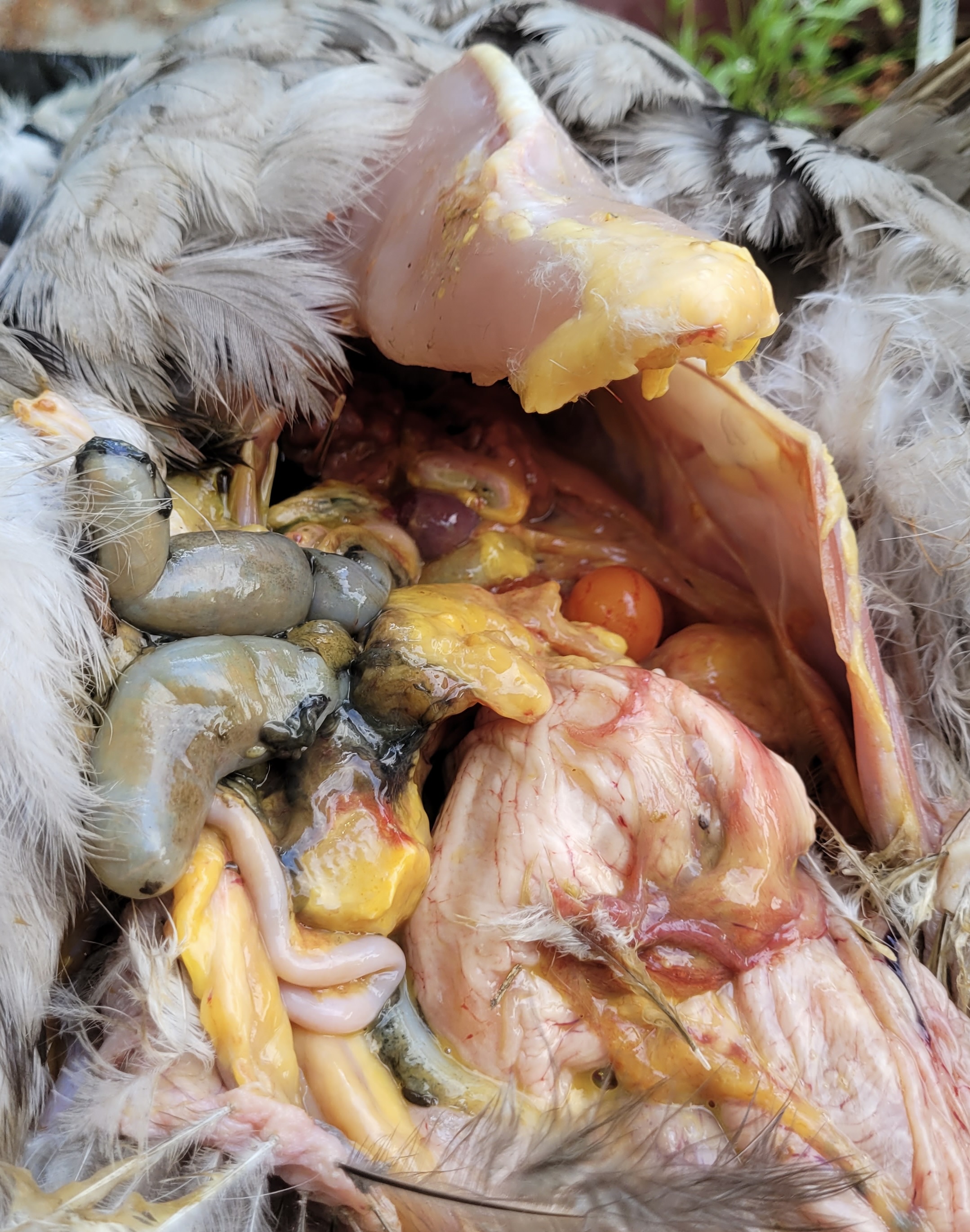
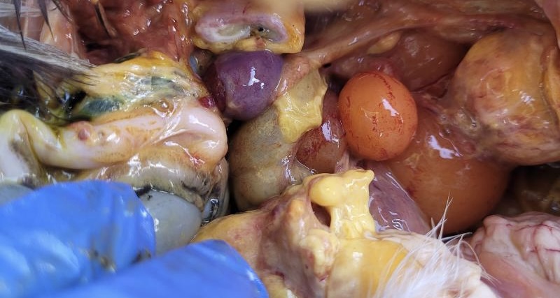
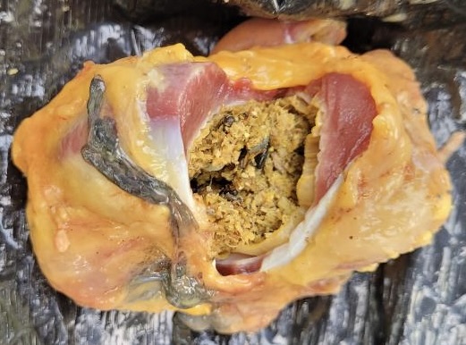
Developing egg yolks; bisected gizzard
The next part was tricky. You know the saying ‘Many hands make light work’ – well I only had two when I really needed four. I wanted to photograph as much as I could, but that meant picking up my cellphone and swiping the screen after each step of the procedure. My gloves became increasingly more gross as they got covered with the infection.
I was not happy to see two large masses at the base of her abdomen. I opened them up and at first glance I already had the answer. Her abdomen and reproductive tract were full of a copious amount of salpingitis (‘lash egg’). I marvelled at how she was still eating, drinking and walking around with that enormous infection brewing in her.
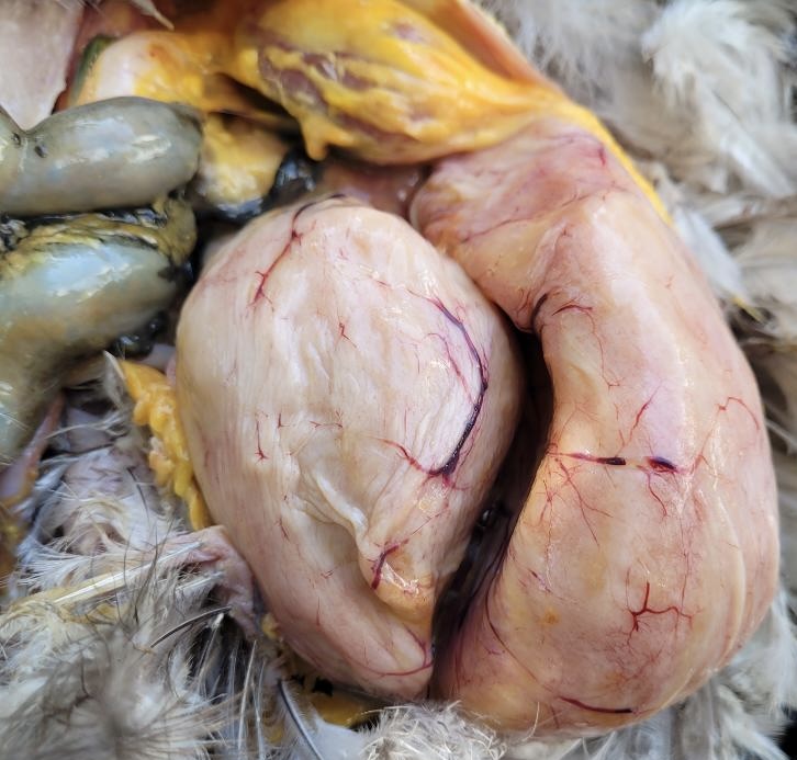
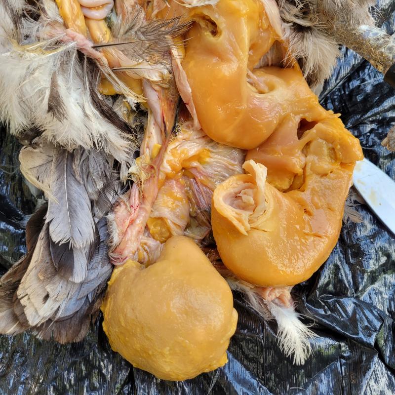
Although she had been dead for less than 12 hours, she smelled. I was working hunched over her while the pungent odour of the infection hit me. I suppose if I did this as a full-time job I’d get used to it but I had to admit it was not pleasant.
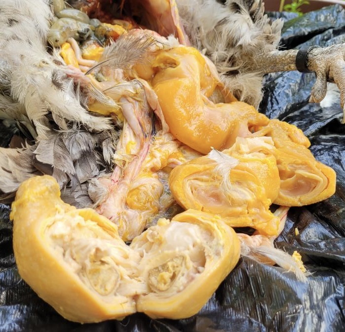
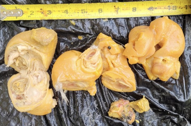
I was curious about the weight of the infection so used my kitchen scale (don’t tell my partner): a whopping 1.4 lbs! Even when that was all removed she was still a decent weight (didn’t get that done).
Clearly this post isn’t about sharing my expertise conducting either humane euthanasia or DIY necropsy. Rather it’s an encouragement to do what you can to take care of your flock around end-of-life issues as well as screw up the courage to do whatever kind of DIY necropsy you’re able in the hope that someone in chickenland can later help with a diagnosis.
“Because chicken science is cooler than you think.”


0 comments on “Case Study: Salpingitis 3”