This series is the result of my partnership with Dr Vicki Bowes, avian vet/pathologist whom I get together with regularly to look at what she calls ‘best guess’ or ‘show and tell’. I present my flash drive full of interesting health issues I’ve collected and we pore over them trying to figure out the most likely diagnosis. Our last meet up resulted in 42 cases. I’ve presented some as a mixed bag and others relating to specific areas of the body (i.e. the head, and legs and feet). This one deals with reproductive tract issues.
“This hen has had water belly for awhile now. I have drained the fluid several times which was a clearish yellow colour. This time it looked like this. I know she has a terminal condition and I am just temporarily fixing the problem, but why is it this colour? I tried three different spots thinking maybe I just got some blood in there, but all spots look exactly the same.” – Dana Zimmerman


Dr Bowes: The dark colour is a sign of an internal hemorrhage. Ascites can be caused by different conditions. If it is related to congestive heart failure the fluid backs up into the liver and leaks into the abdomen. It can also be a sign of ovarian adenocarcinoma. When you drain fluid always make sure the bird is in a standing position and if possible push their intestines up and insert your needle into the abdomen below that. Turning a bird on its back for this procedure can potentially drown them if their air sacs and lungs are compromised. Note: never drain more than 10ml at a time.
“My one year old hen laid regularly, stopped for one week and then laid this weird egg – hard shell with hard, crumbling deposits.” – Janet Bost
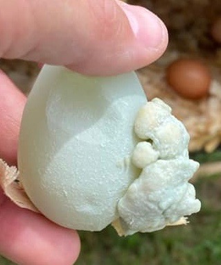
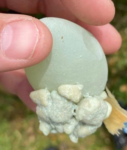
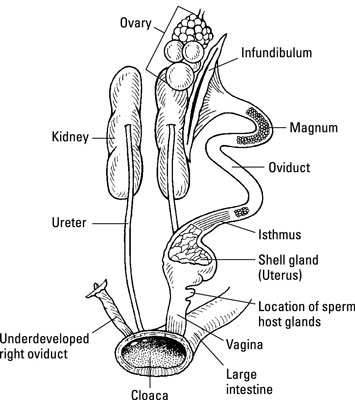
Dr Bowes: This is a case of an abnormally large calcium deposit attached to the egg. Think of an egg developing as though it was on a lathe, slowly turning and adding the various components (i.e. yolk, albumin, membrane, shell, bloom) as it turns and makes it way through the oviduct. The calcium deposit was made in the shell gland and then attached to the eggshell at the end of the production line.
Excess calcium, found in the form of small bumps on the eggshell, can be a result of a hen not fully utilizing all the calcium in her feed intake. There is also a genetic component that causes some hens to lay eggs with calcium deposits (although not usually this size).
Jyn Meyer sent me a photo of this egg with what looks like a spiral inside a watery yolk.
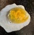
Dr Bowes: Even though it appears yellow this is a yolkless egg. It looks like some reproductive tissue got incorporated into the egg production and like a pearl the other components got wrapped around it. The oviduct was triggered to recognize it and build an egg around it.
My six year old hen quit laying awhile ago and only showed signs of illness the week before she died. When I did a DIY necropsy most of her organs looked fine on initial inspection. I found a softball sized mass on her oviduct which felt solid when I cut into it. There were no signs of egg material or the usual normal infectious material you’d expect to find, just one solid mass. – Debra Watt
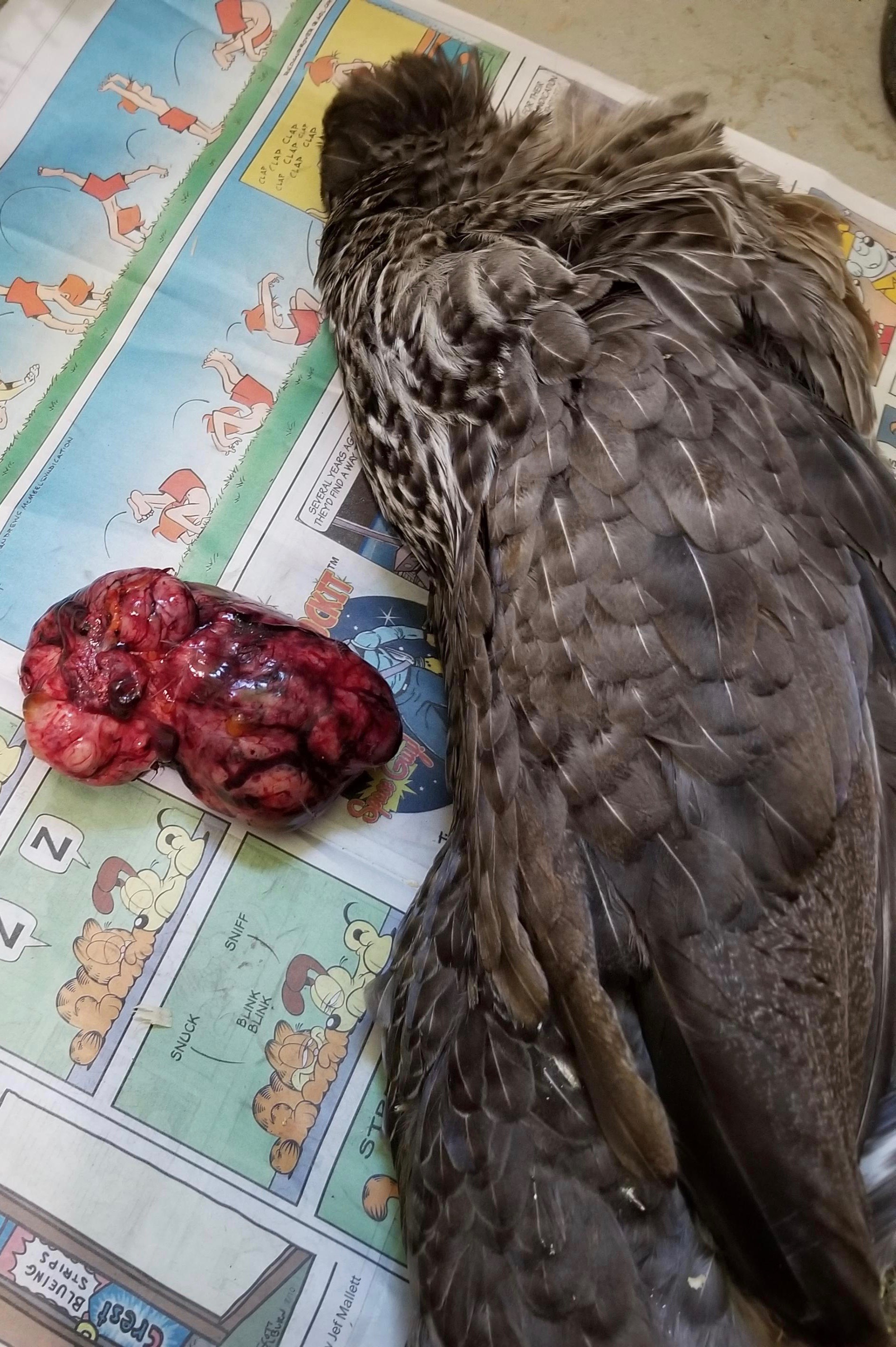

Dr Bowes: I believe this was a case of oviductal adenocarcinoma.
Salpingitis/Oviduct Infection
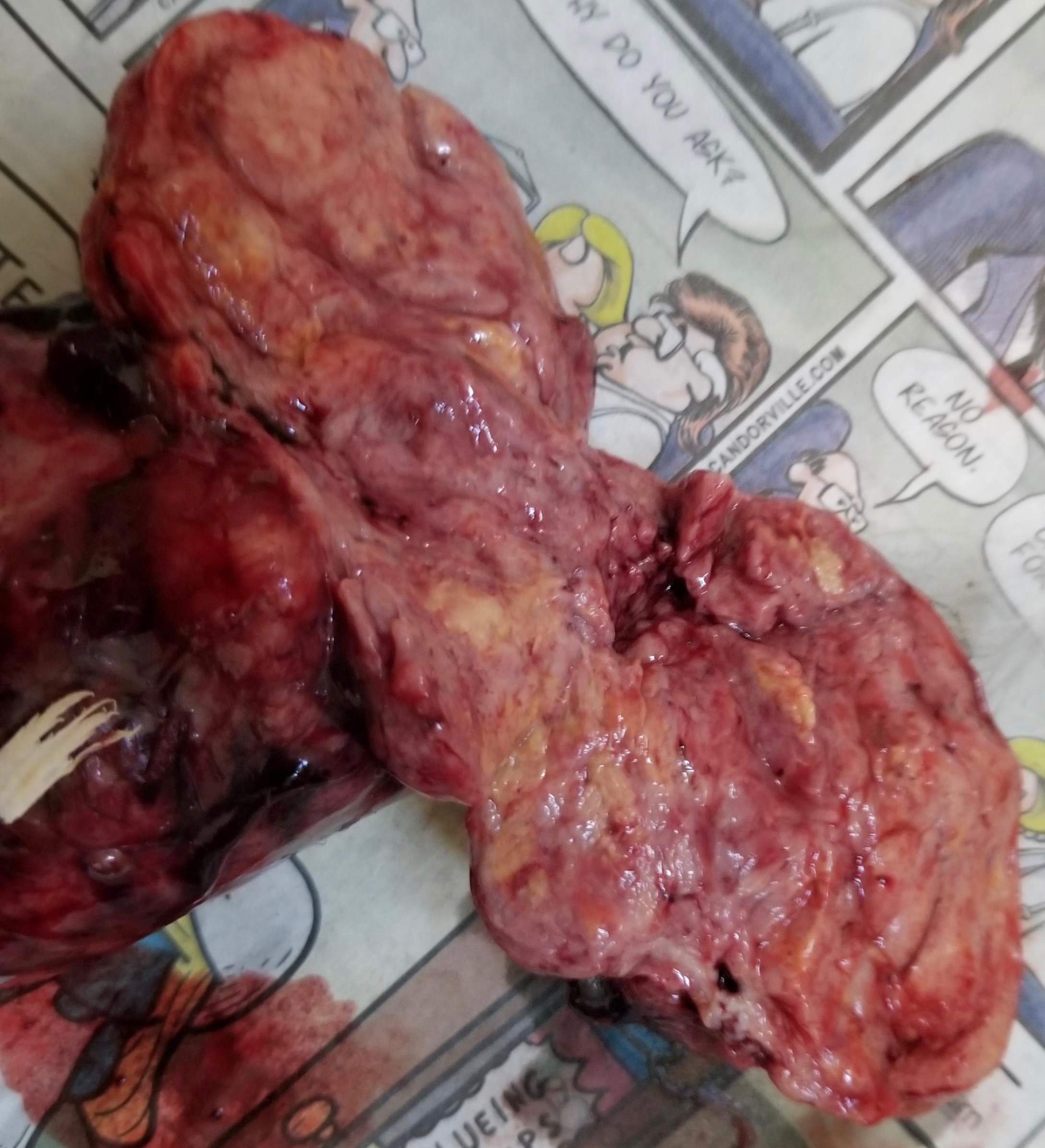
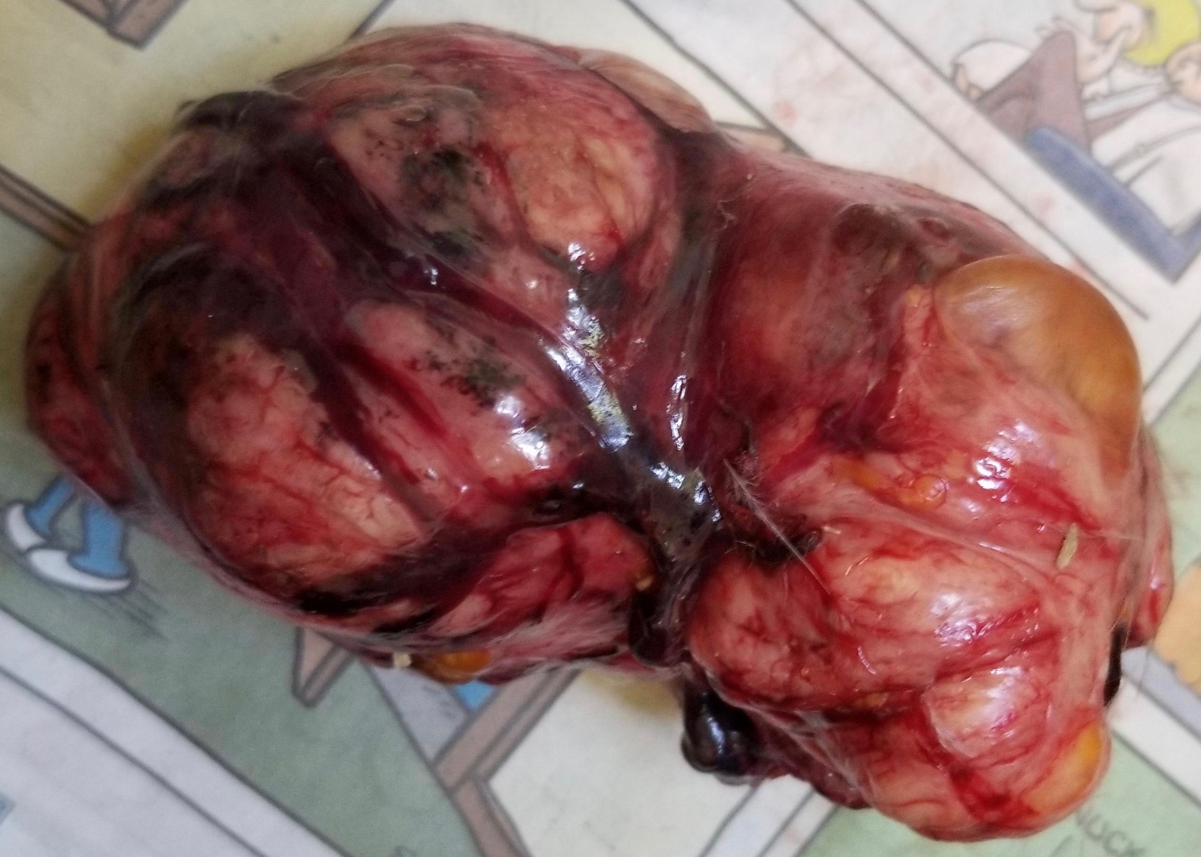
My seven year old hen presented with lethargy, weight loss and a distended abdomen. She had recently dealt with a flystrike infection. I did a DIY necropsy and found what looked like grouping of lash egg clumped together in one big cluster. Her intestines were tight up against her body cavity, and her heart, liver and gizzard all looked remarkably good for her age. She had a huge hard mass in the lower abdomen. – Elizabeth Anderson


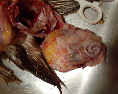


Dr Bowes: This hen had a massively distended abdomen and prominent keel. The weight of the infection meant that the bird can feel a normal weight but was actually emaciated. The infection was encompassed in the oviduct. The hen continued to attempt to lay eggs which contributed to the salpingitis mass (lash egg).
“What can be done to treat this hen?” – Gloria Johnson

Dr Bowes: This is a massive prolapse that involves not just the oviduct but the rectum as well. It’s a complete eversion (turned inside out) of both structures. Recommendation: humane euthanasia
Well that wraps up another edition of Show & Tell With Bitchin’ Chickens and Dr Bowes. I hope that it’s been a learning experience for you.
If you’d like help with a case drop me a line using the ‘contact’ button on my home page. Remember to wear gloves, take good close up photos from several angles and supply us with plenty of information (e.g. timelines, symptoms, medications, general flock health, etc) so we’re able to more accurately pinpoint what’s going on.
Thanks again to Dr Vicki Bowes for her willingness to share her wealth of knowledge and experience to build capacity and skills in small flock keepers.

0 comments on “Avian Pathology Cases: 13”